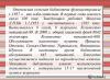
Based on its functional characteristics, the cell membrane can be divided into 9 functions it performs.
Functions of the cell membrane:
1. Transport. Transports substances from cell to cell;
2. Barrier. Has selective permeability, ensures the necessary metabolism;
3. Receptor. Some proteins found in the membrane are receptors;
4. Mechanical. Ensures the autonomy of the cell and its mechanical structures;
5. Matrix. Ensures optimal interaction and orientation of matrix proteins;
6. Energy. Membranes contain energy transfer systems during cellular respiration in mitochondria;
7. Enzymatic. Membrane proteins are sometimes enzymes. For example, intestinal cell membranes;
8. Marking. The membrane contains antigens (glycoproteins) that allow cell identification;
9. Generating. Carries out the generation and conduction of biopotentials.
You can see what a cell membrane looks like using the example of the structure of an animal cell or plant cell.
The figure shows the structure of the cell membrane.
The components of the cell membrane include various cell membrane proteins (globular, peripheral, surface), as well as cell membrane lipids (glycolipid, phospholipid). Also in the structure of the cell membrane there are carbohydrates, cholesterol, glycoprotein and protein alpha helix.
Cell membrane composition
The main composition of the cell membrane includes:
1. Proteins - responsible for various properties of the membrane;
2. Three types of lipids (phospholipids, glycolipids and cholesterol) responsible for membrane rigidity.
Cell membrane proteins:
1. Globular protein;
2. Surface protein;
3. Peripheral protein.
The main purpose of the cell membrane
The main purpose of the cell membrane:
1. Regulate the exchange between the cell and the environment;
2. Separate the contents of any cell from the external environment, thereby ensuring its integrity;
3. Intracellular membranes divide the cell into specialized closed compartments - organelles or compartments in which certain environmental conditions are maintained.
Cell membrane structure
The structure of the cell membrane is a two-dimensional solution of globular integral proteins dissolved in a liquid phospholipid matrix. This model of membrane structure was proposed by two scientists Nicholson and Singer in 1972. Thus, the basis of the membranes is a bimolecular lipid layer, with an ordered arrangement of molecules, as you could see in.
The outside of the cell is covered with a plasma membrane (or outer cell membrane) about 6-10 nm thick.
The cell membrane is a dense film of proteins and lipids (mainly phospholipids). Lipid molecules are arranged in an orderly manner - perpendicular to the surface, in two layers, so that their parts that interact intensively with water (hydrophilic) are directed outward, and their parts inert to water (hydrophobic) are directed inward.
Protein molecules are located in a non-continuous layer on the surface of the lipid framework on both sides. Some of them are immersed in the lipid layer, and some pass through it, forming areas permeable to water. These proteins perform various functions - some of them are enzymes, others are transport proteins involved in the transfer of certain substances from the environment to the cytoplasm and in the opposite direction.
Basic functions of the cell membrane
One of the main properties of biological membranes is selective permeability (semi-permeability)- some substances pass through them with difficulty, others easily and even towards higher concentrations. Thus, for most cells, the concentration of Na ions inside is significantly lower than in the environment. The opposite relationship is typical for K ions: their concentration inside the cell is higher than outside. Therefore, Na ions always tend to penetrate the cell, and K ions always tend to exit. The equalization of the concentrations of these ions is prevented by the presence in the membrane of a special system that plays the role of a pump, which pumps Na ions out of the cell and simultaneously pumps K ions inside.
The tendency of Na ions to move from outside to inside is used to transport sugars and amino acids into the cell. With the active removal of Na ions from the cell, conditions are created for the entry of glucose and amino acids into it.
In many cells, substances are also absorbed by phagocytosis and pinocytosis. At phagocytosis the flexible outer membrane forms a small depression into which the captured particle falls. This recess increases, and, surrounded by a section of the outer membrane, the particle is immersed in the cytoplasm of the cell. The phenomenon of phagocytosis is characteristic of amoebas and some other protozoa, as well as leukocytes (phagocytes). Cells absorb liquids containing substances necessary for the cell in a similar way. This phenomenon was called pinocytosis.
The outer membranes of different cells differ significantly both in the chemical composition of their proteins and lipids, and in their relative content. It is these features that determine the diversity in the physiological activity of the membranes of various cells and their role in the life of cells and tissues.
The endoplasmic reticulum of the cell is connected to the outer membrane. With the help of outer membranes, various types of intercellular contacts are carried out, i.e. communication between individual cells.
Many types of cells are characterized by the presence on their surface of a large number of protrusions, folds, and microvilli. They contribute to both a significant increase in cell surface area and improved metabolism, as well as stronger connections between individual cells and each other.
Plant cells have thick membranes on the outside of the cell membrane, clearly visible under an optical microscope, consisting of fiber (cellulose). They create a strong support for plant tissues (wood).
Some animal cells also have a number of external structures located on top of the cell membrane and have a protective nature. An example is the chitin of insect integumentary cells.
Functions of the cell membrane (briefly)
| Function | Description |
|---|---|
| Protective Barrier | Separates internal cell organelles from the external environment |
| Regulatory | Regulates the metabolism between the internal contents of the cell and the external environment |
| Delimiting (compartmentalization) | Division of the internal space of the cell into independent blocks (compartments) |
| Energy | - Energy accumulation and transformation; - light reactions of photosynthesis in chloroplasts; - Absorption and secretion. |
| Receptor (informational) | Participates in the formation of arousal and its conduct. |
| Motor | Carries out the movement of the cell or its individual parts. |
The cell membrane is called plasmalemma or plasma membrane. The main functions of the cell membrane are maintaining the integrity of the cell and interconnecting with the external environment.
Structure
Cell membranes consist of lipoprotein (fat-protein) structures and have a thickness of 10 nm. The membrane walls are formed by three classes of lipids:
- phospholipids - compounds of phosphorus and fats;
- glycolipids - compounds of lipids and carbohydrates;
- cholesterol (cholesterol) - fatty alcohol.
These substances form a liquid mosaic structure consisting of three layers. Phospholipids form the two outer layers. They have a hydrophilic head from which two hydrophobic tails extend. The tails are turned inside the structure, forming an inner layer. When cholesterol is incorporated into the phospholipid tails, the membrane becomes rigid.

Rice. 1. Membrane structure.
Built between the phospholipids are glycolipids that perform a receptor function and two types of proteins:
- peripheral (external, superficial) - located on the lipid surface, without penetrating deep into the membrane;
- integral - embedded at different levels, can penetrate the entire membrane, only the inner or outer lipid layer;
All proteins differ in their structure and perform different functions. For example, globular protein compounds have a hydrophobic-hydrophilic structure and perform a transport function.
TOP 4 articleswho are reading along with this

Rice. 2. Types of membrane proteins.
Plasmalemma is a fluid structure, because the lipids are not interconnected, but are simply arranged in dense rows. Thanks to this property, the membrane can change configuration, be mobile and elastic, and also transport substances.
Functions
What functions does the cell membrane perform?
- barrier - separates the contents of the cell from the external environment;
- transport - regulates metabolism;
- enzymatic - carries out enzymatic reactions;
- receptor - recognizes external stimuli.
The most important function is the transport of substances during metabolism. Liquid and solid substances constantly enter the cell from the external environment. Metabolic products come out. All substances pass through the cell membrane. Transport occurs in several ways, which are described in the table.
|
View |
Substances |
Process |
|
Diffusion |
Gases, fat-soluble molecules |
Uncharged molecules pass through the lipid layer freely or with the help of a special protein channel without expending energy |
|
Solutions |
One-way diffusion towards higher solute concentration |
|
|
Endocytosis |
Solid and liquid substances of the external environment |
The transfer of liquids is called pinocytosis, and the transfer of solids is called phagocytosis. Penetrate by pulling the membrane inward until a bubble forms |
|
Exocytosis |
Solid and liquid substances of the internal environment |
The reverse process of endocytosis. Bubbles containing substances are moved by the cytoplasm to the membrane and merge with it, releasing the contents out |

Rice. 3. Endocytosis and exocytosis.
Active transport of substance molecules (sodium-potassium pump) is carried out using protein structures built into the membrane and requires energy in the form of ATP.
Average rating: 4.7. Total ratings received: 289.
Outer cell membrane (plasmalemma, cytolemma, plasma membrane) of animal cells covered on the outside (i.e., on the side not in contact with the cytoplasm) with a layer of oligosaccharide chains covalently attached to membrane proteins (glycoproteins) and to a lesser extent to lipids (glycolipids). This carbohydrate membrane coating is called glycocalyx. The purpose of the glycocalyx is not yet very clear; there is an assumption that this structure takes part in the processes of intercellular recognition.
In plant cells On top of the outer cell membrane there is a dense cellulose layer with pores, through which communication between neighboring cells occurs through cytoplasmic bridges.
In cells mushrooms on top of the plasmalemma - a dense layer chitin.
U bacteria – mureina.
Properties of biological membranes
1. Self-assembly ability after destructive influences. This property is determined by the physicochemical properties of phospholipid molecules, which in an aqueous solution come together so that the hydrophilic ends of the molecules unfold outward, and the hydrophobic ends inward. Proteins can be built into ready-made phospholipid layers. The ability to self-assemble is important at the cellular level.
2. Semi-permeable(selectivity in the transmission of ions and molecules). Ensures the maintenance of constancy of the ionic and molecular composition in the cell.
3. Membrane fluidity. Membranes are not rigid structures; they constantly fluctuate due to the rotational and vibrational movements of lipid and protein molecules. This ensures a higher rate of enzymatic and other chemical processes in membranes.
4. Membrane fragments do not have free ends, as they close into bubbles.
Functions of the outer cell membrane (plasmalemma)
The main functions of the plasmalemma are the following: 1) barrier, 2) receptor, 3) exchange, 4) transport.
1. Barrier function. It is expressed in the fact that the plasmalemma limits the contents of the cell, separating it from the external environment, and intracellular membranes divide the cytoplasm into separate reaction cells. compartments.
2. Receptor function. One of the most important functions of the plasmalemma is to ensure communication (connection) of the cell with the external environment through the receptor apparatus present in the membranes, which is of a protein or glycoprotein nature. The main function of the receptor formations of the plasmalemma is the recognition of external signals, thanks to which cells are correctly oriented and form tissues during the process of differentiation. The receptor function is associated with the activity of various regulatory systems, as well as the formation of an immune response.
Exchange function determined by the content of enzyme proteins in biological membranes, which are biological catalysts. Their activity varies depending on the pH of the environment, temperature, pressure, and the concentration of both the substrate and the enzyme itself. Enzymes determine the intensity of key reactions metabolism, as well as their direction.
Transport function of membranes. The membrane allows for selective penetration of various chemicals into the cell and out of the cell into the environment. Transport of substances is necessary to maintain the appropriate pH and proper ionic concentration in the cell, which ensures the efficiency of cellular enzymes. Transport supplies nutrients that serve as a source of energy as well as material for the formation of various cellular components. The removal of toxic waste from the cell, the secretion of various useful substances and the creation of ion gradients necessary for nervous and muscle activity depend on it. Changes in the rate of transfer of substances can lead to disturbances in bioenergetic processes, water-salt metabolism, excitability and other processes. Correction of these changes underlies the action of many medications.
There are two main ways for substances to enter the cell and exit the cell into the external environment;
passive transport,
active transport.
Passive transport follows a chemical or electrochemical concentration gradient without the expenditure of ATP energy. If the molecule of the transported substance has no charge, then the direction of passive transport is determined only by the difference in the concentration of this substance on both sides of the membrane (chemical concentration gradient). If the molecule is charged, then its transport is affected by both the chemical concentration gradient and the electrical gradient (membrane potential).
Both gradients together constitute the electrochemical gradient. Passive transport of substances can be carried out in two ways: simple diffusion and facilitated diffusion.
With simple diffusion salt ions and water can penetrate through selective channels. These channels are formed by certain transmembrane proteins that form end-to-end transport pathways that are open permanently or only for a short time. Various molecules of size and charge corresponding to the channels penetrate through selective channels.
There is another way of simple diffusion - this is the diffusion of substances through the lipid bilayer, through which fat-soluble substances and water easily pass. The lipid bilayer is impermeable to charged molecules (ions), and at the same time, uncharged small molecules can diffuse freely, and the smaller the molecule, the faster it is transported. The rather high rate of diffusion of water through the lipid bilayer is precisely explained by the small size of its molecules and the lack of charge.
With facilitated diffusion Transport of substances involves proteins - carriers that work on the “ping-pong” principle. In this case, the protein exists in two conformational states: in the “pong” state, the binding sites for the transported substance are open on the outside of the bilayer, and in the “ping” state, the same sites are open on the other side. This process is reversible. From which side the binding site of a substance will be open at a given time depends on the concentration gradient of this substance.
In this way, sugars and amino acids pass through the membrane.
With facilitated diffusion, the rate of transport of substances increases significantly compared to simple diffusion.
In addition to carrier proteins, some antibiotics are involved in facilitated diffusion, for example, gramicidin and valinomycin.
Because they provide ion transport, they are called ionophores.
Active transport of substances in the cell. This type of transport always costs energy. The source of energy required for active transport is ATP. A characteristic feature of this type of transport is that it is carried out in two ways:
using enzymes called ATPases;
transport in membrane packaging (endocytosis).
IN The outer cell membrane contains enzyme proteins such as ATPases, whose function is to provide active transport ions against a concentration gradient. Since they provide ion transport, this process is called an ion pump.
There are four main known ion transport systems in animal cells. Three of them provide transfer through biological membranes: Na + and K +, Ca +, H +, and the fourth - transfer of protons during the functioning of the mitochondrial respiratory chain.
An example of an active ion transport mechanism is sodium-potassium pump in animal cells. It maintains a constant concentration of sodium and potassium ions in the cell, which differs from the concentration of these substances in the environment: normally, there are fewer sodium ions in the cell than in the environment, and more potassium ions.
As a result, according to the laws of simple diffusion, potassium tends to leave the cell, and sodium diffuses into the cell. In contrast to simple diffusion, the sodium-potassium pump constantly pumps sodium out of the cell and introduces potassium: for every three molecules of sodium released out, there are two molecules of potassium introduced into the cell.
This transport of sodium-potassium ions is ensured by the dependent ATPase, an enzyme localized in the membrane in such a way that it penetrates its entire thickness. Sodium and ATP enter this enzyme from the inside of the membrane, and potassium from the outside.
The transfer of sodium and potassium across the membrane occurs as a result of conformational changes that the sodium-potassium dependent ATPase undergoes, which is activated when the concentration of sodium inside the cell or potassium in the environment increases.
To supply energy to this pump, ATP hydrolysis is necessary. This process is ensured by the same enzyme, sodium-potassium dependent ATPase. Moreover, more than one third of the ATP consumed by an animal cell at rest is spent on the operation of the sodium-potassium pump.
Violation of the proper functioning of the sodium-potassium pump leads to various serious diseases.
The efficiency of this pump exceeds 50%, which is not achieved by the most advanced machines created by man.
Many active transport systems are powered by energy stored in ion gradients rather than by direct hydrolysis of ATP. All of them work as cotransport systems (promoting the transport of low molecular weight compounds). For example, the active transport of some sugars and amino acids into animal cells is determined by a sodium ion gradient, and the higher the sodium ion gradient, the greater the rate of glucose absorption. And, conversely, if the sodium concentration in the intercellular space decreases markedly, glucose transport stops. In this case, sodium must join the sodium-dependent glucose transport protein, which has two binding sites: one for glucose, the other for sodium. Sodium ions penetrating the cell facilitate the introduction of the carrier protein into the cell along with glucose. Sodium ions that enter the cell along with glucose are pumped back by sodium-potassium dependent ATPase, which, by maintaining a sodium concentration gradient, indirectly controls glucose transport.
Transport of substances in membrane packaging. Large molecules of biopolymers practically cannot penetrate through the plasmalemma by any of the above-described mechanisms of transport of substances into the cell. They are captured by the cell and absorbed into membrane packaging, which is called endocytosis. The latter is formally divided into phagocytosis and pinocytosis. The uptake of particulate matter by the cell is phagocytosis, and liquid - pinocytosis. During endocytosis, the following stages are observed:
reception of the absorbed substance due to receptors in the cell membrane;
invagination of the membrane with the formation of a bubble (vesicle);
separation of the endocytic vesicle from the membrane with energy consumption – phagosome formation and restoration of membrane integrity;
Fusion of the phagosome with the lysosome and formation phagolysosomes (digestive vacuole) in which digestion of absorbed particles occurs;
removal of material undigested in the phagolysosome from the cell ( exocytosis).
In the animal world endocytosis is a characteristic method of nutrition of many unicellular organisms (for example, in amoebas), and among many cellular organisms, this type of digestion of food particles is found in the endodermal cells of coelenterates. As for mammals and humans, they have a reticulo-histio-endothelial system of cells with the ability to endocytosis. Examples include blood leukocytes and liver Kupffer cells. The latter line the so-called sinusoidal capillaries of the liver and capture various foreign particles suspended in the blood. Exocytosis- This is also a method of removing from the cell of a multicellular organism the substrate secreted by it, which is necessary for the function of other cells, tissues and organs.
The membrane is an ultra-fine structure that forms the surfaces of organelles and the cell as a whole. All membranes have a similar structure and are connected into one system.
Chemical composition
Cell membranes are chemically homogeneous and consist of proteins and lipids of various groups:
- phospholipids;
- galactolipids;
- sulfolipids.
They also contain nucleic acids, polysaccharides and other substances.
Physical properties
At normal temperatures, the membranes are in a liquid crystalline state and constantly fluctuate. Their viscosity is close to that of vegetable oil.
The membrane is recoverable, durable, elastic and porous. Membrane thickness is 7 - 14 nm.
TOP 4 articleswho are reading along with this
The membrane is impermeable to large molecules. Small molecules and ions can pass through the pores and the membrane itself under the influence of concentration differences on different sides of the membrane, as well as with the help of transport proteins.
Model
Typically, the structure of membranes is described using a fluid mosaic model. The membrane has a frame - two rows of lipid molecules, tightly adjacent to each other, like bricks.

Rice. 1. Sandwich-type biological membrane.
On both sides, the surface of lipids is covered with proteins. The mosaic pattern is formed by protein molecules unevenly distributed on the surface of the membrane.
According to the degree of immersion in the bilipid layer, protein molecules are divided into three groups:
- transmembrane;
- submerged;
- superficial.
Proteins provide the main property of the membrane - its selective permeability to various substances.
Membrane types
All cell membranes according to localization can be divided into the following types:
- external;
- nuclear;
- organelle membranes.
The outer cytoplasmic membrane, or plasmolemma, is the boundary of the cell. Connecting with the elements of the cytoskeleton, it maintains its shape and size.

Rice. 2. Cytoskeleton.
The nuclear membrane, or karyolemma, is the boundary of the nuclear contents. It is constructed of two membranes, very similar to the outer one. The outer membrane of the nucleus is connected to the membranes of the endoplasmic reticulum (ER) and, through pores, to the inner membrane.
ER membranes penetrate the entire cytoplasm, forming surfaces on which the synthesis of various substances, including membrane proteins, occurs.
Organelle membranes
Most organelles have a membrane structure.
The walls are built from one membrane:
- Golgi complex;
- vacuoles;
- lysosomes
Plastids and mitochondria are built from two layers of membranes. Their outer membrane is smooth, and the inner one forms many folds.
Features of photosynthetic membranes of chloroplasts are built-in chlorophyll molecules.
Animal cells have a carbohydrate layer on the surface of their outer membrane called the glycocalyx.

Rice. 3. Glycocalyx.
The glycocalyx is most developed in the cells of the intestinal epithelium, where it creates conditions for digestion and protects the plasmalemma.
Table "Structure of the cell membrane"
What have we learned?
We looked at the structure and functions of the cell membrane. The membrane is a selective (selective) barrier of the cell, nucleus and organelles. The structure of the cell membrane is described by the fluid mosaic model. According to this model, protein molecules are built into the bilayer of viscous lipids.
Test on the topic
Evaluation of the report
Average rating: 4.5. Total ratings received: 270.