Nucleic acids are high-molecular substances consisting of mononucleotides, which are connected to each other in a polymer chain using 3", 5" phosphodiester bonds and are packaged in cells in a certain way.
Nucleic acids are biopolymers of two types: ribonucleic acid (RNA) and deoxyribonucleic acid (DNA). Each biopolymer consists of nucleotides that differ in the carbohydrate residue (ribose, deoxyribose) and one of the nitrogenous bases (uracil, thymine). According to these differences, nucleic acids received their name.
Structure of ribonucleic acid
Primary structure of RNA
RNA molecule are linear (i.e., unbranched) polynucleotides with a similar principle of organization to DNA. RNA monomers are nucleotides consisting of phosphoric acid, a carbohydrate (ribose) and a nitrogenous base, connected by 3", 5" phosphodiester bonds. The polynucleotide chains of the RNA molecule are polar, i.e. have distinguishable 5' and 3" ends. Moreover, unlike DNA, RNA is a single-stranded molecule. The reason for this difference is three features of the primary structure:- RNA, unlike DNA, contains ribose instead of deoxyribose, which has an additional hydroxy group. The hydroxy group makes the double-chain structure less compact
- Among the four main, or major, nitrogenous bases (A, G, C and U), instead of thymine, uracil is contained, which differs from thymine only in the absence of a methyl group in the 5th position. Due to this, the strength of the hydrophobic interaction in the complementary A-U pair decreases, which also reduces the likelihood of the formation of stable double-chain molecules.
- Finally, RNA (especially tRNA) has a high content of the so-called. minor bases and nucleosides. Among them are dihydrouridine (uracil does not have one double bond), pseudouridine (uracil is associated with ribose differently than usual), dimethyladenine and dimethylguanine (in nitrogenous bases there are two additional methyl groups) and many others. Almost all of these bases cannot participate in complementary interactions. Thus, methyl groups in dimethyladenine (unlike thymine and 5-methylcytosine) are located at an atom that forms a hydrogen bond in the A-U pair; therefore, now this connection cannot be closed. This also prevents the formation of double-stranded molecules.
Thus, the well-known differences in the composition of RNA from DNA are of great biological importance: after all, RNA molecules can perform their function only in a single-stranded state, which is most obvious for mRNA: it is difficult to imagine how a double-stranded molecule could be translated on ribosomes.
At the same time, while remaining single, in some areas the RNA chain can form loops, protrusions or “hairpins” with a double-stranded structure (Fig. 1). This structure is stabilized by the interaction of bases in pairs A::U and G:::C. However, “incorrect” pairs can also form (for example, G U), and in some places there are “hairpins” and no interaction occurs at all. Such loops can contain (especially in tRNA and rRNA) up to 50% of all nucleotides. The total content of nucleotides in RNA varies from 75 units to many thousands. But even the largest RNAs are several orders of magnitude shorter than chromosomal DNA.
The primary structure of mRNA is copied from a section of DNA containing information about the primary structure of the polypeptide chain. The primary structure of other types of RNA (tRNA, rRNA, rare RNA) is the final copy of the genetic program of the corresponding DNA genes.
Secondary and tertiary structures of RNA
Ribonucleic acids (RNA) are single-stranded molecules, so, unlike DNA, their secondary and tertiary structures are irregular. These structures, defined as the spatial conformation of a polynucleotide chain, are formed mainly by hydrogen bonds and hydrophobic interactions between nitrogenous bases. If the native DNA molecule is characterized by a stable helix, then the structure of RNA is more diverse and labile. X-ray diffraction analysis showed that individual sections of the RNA polynucleotide chain, bending, wind on themselves to form intrahelical structures. Stabilization of structures is achieved through complementary pairing of nitrogenous bases of antiparallel sections of the chain; the specific pairs here are A-U, G-C and, less commonly, G-U. Due to this, both short and extended double-helical regions appear in the RNA molecule, belonging to the same chain; these areas are called hairpins. The model of RNA secondary structure with hairpin elements was created in the late 50s - early 60s. XX century in the laboratories of A. S. Spirin (Russia) and P. Doty (USA).
| Some types of RNA | ||
| Types of RNA | Size in nucleotides | Function |
| gRNA - genomic RNA | 10000-100000 | |
| mRNA - informational (matrix) RNA | 100-100000 | transmits information about protein structure from a DNA molecule |
| tRNA - transfer RNA | 70-90 | transports amino acids to the site of protein synthesis |
| rRNA - ribosomal RNA | several discrete classes from 100 to 500,000 | found in ribosomes, participates in maintaining the structure of the ribosome |
| sn-RNA - small nuclear RNA | 100 | removes introns and enzymatically joins exons in mRNA |
| sno-RNA - small nucleolar RNA | is involved in directing or carrying out base modifications in rRNA and small nuclear RNA, such as methylation and pseudouridination. Most small nucleolar RNAs are found in introns of other genes | |
| srp-RNA - signal recognition RNA | recognizes the signal sequence of proteins intended for expression and participates in their transport across the cytoplasmic membrane | |
| mi-RNA - micro-RNA | 22 | control the translation of structural genes by complementary binding to the 3" ends of untranslated regions of mRNA |
The formation of helical structures is accompanied by a hypochromic effect - a decrease in the optical density of RNA samples at 260 nm. The destruction of these structures occurs when the ionic strength of the RNA solution is reduced or when it is heated to 60-70 ° C; it is also called melting and is explained by the structural transition of a helix - a chaotic coil, which is accompanied by an increase in the optical density of the nucleic acid solution.
There are several types of RNA in cells:
- information (or messenger) RNA (mRNA or mRNA) and its predecessor - heterogeneous nuclear RNA (r-n-RNA)
- transfer RNA (tRNA) and its precursor
- ribosomal (rRNA) and its precursor
- small nuclear RNA (sn-RNA)
- small nucleolar RNA (sno-RNA)
- signal recognition RNA (srp-RNA)
- micro-RNA (mi-RNA)
- mitochondrial RNA (t+ RNA).

Heterogeneous nuclear and messenger RNA
Heterogeneous nuclear RNA is characteristic exclusively of eukaryotes. It is the precursor of messenger RNA (mRNA), which carries genetic information from nuclear DNA to the cytoplasm. Heterogeneous nuclear RNA (pre-mRNA) was discovered by the Soviet biochemist G. P. Georgiev. The number of types of r-RNA is equal to the number of genes, since it serves as a direct copy of the coding sequences of the genome, due to which it has copies of DNA palindromes, therefore its secondary structure contains hairpins and linear regions. In the process of transcription of RNA from DNA, the enzyme RNA polymerase II plays a key role.
Messenger RNA is formed as a result of processing (maturation) of r-RNA, during which hairpins are cut off, non-coding regions (introns) are excised, and coding exons are glued together.
Messenger RNA (i-RNA) is a copy of a specific section of DNA and acts as a carrier of genetic information from DNA to the site of protein synthesis (ribosomes) and is directly involved in the assembly of its molecules.
Mature messenger RNA has several regions with different functional roles (Fig.)
- at the 5" end there is a so-called "cap" or cap - a section of one to four modified nucleotides. This structure protects the 5" end of the mRNA from endonucleases
- after the “cap” there is a 5"-untranslated region - a sequence of several dozen nucleotides. It is complementary to one of the sections of the r-RNA that is part of the small subunit of the ribosome. Due to this, it serves for the primary binding of m-RNA to the ribosome, but itself not broadcast
- the initiation codon is AUG, encoding methionine. All mRNAs have the same start codon. Translation (reading) of m-RNA begins with it. If methionine is not needed after synthesis of the peptide chain, then it is usually cleaved from its N-terminus.
- The start codon is followed by a coding portion, which contains information about the amino acid sequence of the protein. In eukaryotes, mature m-RNAs are monocistronic, i.e. each of them carries information about the structure of only one polypeptide chain.
Another thing is that sometimes the peptide chain, soon after formation on the ribosome, is cut into several smaller chains. This happens, for example, during the synthesis of insulin and a number of oligopeptide hormones.
The coding part of the mature m-RNA of eukaryotes is devoid of introns - any inserted non-coding sequences. In other words, there is a continuous sequence of sense codons that must be read in the direction 5" -> 3".
- At the end of this sequence there is a termination codon - one of three “senseless” codons: UAA, UAG or UGA (see genetic code table below).
- This codon may be followed by another 3' untranslated region, which is significantly longer than the 5' untranslated region.
- Finally, almost all mature eukaryotic mRNAs (except histone mRNAs) contain a poly(A) fragment of 150-200 adenyl nucleotides at the 3" end
The 3" untranslated region and the poly(A) fragment are related to the regulation of the lifespan of m-RNA, since the destruction of m-RNA is carried out by 3" exonucleases. After the end of m-RNA translation, 10-15 nucleotides are cleaved from the poly(A) fragment. When this fragment is exhausted, a significant part of the mRNA begins to degrade (if the 3" untranslated region is missing).
The total number of nucleotides in mRNA usually varies within several thousand. At the same time, the coding part can sometimes account for only 60-70% of the nucleotides.
In cells, mRNA molecules are almost always associated with proteins. The latter probably stabilize the linear structure of the mRNA, i.e., they prevent the formation of “hairpins” in the coding part. In addition, proteins can protect mRNA from premature destruction. Such complexes of mRNA with proteins are sometimes called informosomes.
Transfer RNA in the cytoplasm of the cell carries amino acids in activated form to ribosomes, where they are combined into peptide chains in a specific sequence, which is specified by the RNA matrix (mRNA). Currently, nucleotide sequence data are known for more than 1,700 tRNA species from prokaryotic and eukaryotic organisms. They all have common features both in their primary structure and in the way the polynucleotide chain is folded into a secondary structure due to the complementary interaction of the nucleotides included in their structure.

Transfer RNA contains no more than 100 nucleotides, among which there is a high content of minor, or modified, nucleotides. The first transfer RNA to be completely deciphered was alanine RNA, isolated from yeast. The analysis showed that alanine RNA consists of 77 nucleotides located in a strictly defined sequence; they contain so-called minor nucleotides, represented by atypical nucleosides
Alanine tRNA contains 9 unusual bases with one or more methyl groups, which are added to them enzymatically after the formation of phosphodiester bonds between the nucleotides. These bases are incapable of forming ordinary pairs; perhaps they serve to prevent base pairing in certain parts of the molecule and thus expose specific chemical groups that form secondary bonds with messenger RNA, a ribosome, or perhaps an enzyme necessary to attach a particular amino acid to the corresponding transfer RNA. The known sequence of nucleotides in a tRNA essentially means that its sequence in the genes on which this tRNA is synthesized is also known. This sequence can be deduced based on the rules for specific base pairing established by Watson and Crick. In 1970, a complete double-stranded DNA molecule with a corresponding sequence of 77 nucleotides was synthesized, and it turned out that it could serve as a template for the construction of alanine transfer RNA. This was the first artificially synthesized gene. |

tRNA transcription
Transcription of tRNA molecules occurs from the sequences encoding it in DNA with the participation of the enzyme RNA polymerase III. During transcription, the primary structure of tRNA is formed in the form of a linear molecule. Formation begins with the compilation of a nucleotide sequence by RNA polymerase in accordance with the gene containing information about this transfer RNA. This sequence is a linear polynucleotide chain in which the nucleotides follow each other. The linear polynucleotide chain is the primary RNA, the predecessor of tRNA, which includes introns - uninformative excess nucleotides. At this level of organization, pre-tRNA is not functional. Formed in different places on the DNA of chromosomes, pre-tRNA contains a surplus of approximately 40 nucleotides compared to the mature tRNA.
The second step is that the newly synthesized tRNA precursor undergoes post-transcriptional maturation or processing. During processing, uninformative excesses in pre-RNA are removed and mature, functional RNA molecules are formed.
Pre-tRNA processing
Processing begins with the formation of intramolecular hydrogen bonds in the transcript and the tRNA molecule takes the shape of a cloverleaf. This is the secondary level of tRNA organization, at which the tRNA molecule is not yet functional. Next, the non-informative sections of the pre-RNA are cut out, the informative sections of the “broken genes” are spliced - splicing and modification of the 5" and 3" terminal sections of the RNA.
Excision of non-informative sections of pre-RNA is carried out using ribonucleases (exo- and endonucleases). After removal of excess nucleotides, tRNA bases are methylated. The reaction is carried out by methyltransferases. S-adenosylmethionine acts as a donor of methyl groups. Methylation prevents the destruction of tRNA by nucleases. The finally mature tRNA is formed by the addition of a specific triple of nucleotides (acceptor end) - CCA, which is carried out by a special RNA polymerase.
Upon completion of processing, additional hydrogen bonds are again formed in the secondary structure, due to which tRNA moves to the tertiary level of organization and takes the form of the so-called L-form. In this form, tRNA enters the hyaloplasm.
Structure of tRNA
The structure of transfer RNA is based on a chain of nucleotides. However, due to the fact that any chain of nucleotides has positively and negatively charged parts, it cannot be in an unfolded state in the cell. These charged parts, being attracted to each other, easily form hydrogen bonds with each other according to the principle of complementarity. Hydrogen bonds intricately twist the t-RNA strand and hold it in this position. As a result, the secondary structure of t-RNA has the appearance of a “clover leaf” (Fig.), containing 4 double-stranded sections in its structure. A high content of minor or modified nucleotides, noted in the tRNA chain and incapable of complementary interactions, forms 5 single-stranded regions.
That. The secondary structure of tRNA is formed as a result of intrastrand pairing of complementary nucleotides of individual sections of tRNA. Regions of tRNA that are not involved in the formation of hydrogen bonds between nucleotides form loops or linear units. The following structural regions are distinguished in tRNA:
- Acceptor site (end), consisting of four linearly arranged nucleotides, three of which have the same sequence in all types of tRNA - CCA. The hydroxyl 3"-OH of adenosine is free. An amino acid is attached to it by the carboxyl group, hence the name of this region of tRNA - acceptor. The tRNA amino acid bound to the 3"-hydroxyl group of adenosine is delivered to the ribosomes, where protein synthesis occurs.
- Anticodon loop, usually formed by seven nucleotides. It contains a triplet of nucleotides specific to each tRNA, called an anticodon. The tRNA anticodon pairs with the mRNA codon according to the principle of complementarity. Codon-anticodon interactions determine the order of amino acids in the polypeptide chain during its assembly in ribosomes.
- Pseudouridyl loop (or TΨC loop), consisting of seven nucleotides and necessarily containing a pseudouridylic acid residue. It is assumed that the pseudouridyl loop is involved in the binding of tRNA to the ribosome.
- Dihydrouridine or D-loop, usually consisting of 8-12 nucleotide residues, among which there are always several dihydrouridine residues. It is believed that the D-loop is necessary for binding to aminoacyl-tRNA synthetase, which is involved in the recognition of its tRNA by an amino acid (see “Protein biosynthesis”),
- Additional loop, which varies in size and nucleotide composition for different tRNAs.
The tertiary structure of tRNA is no longer cloverleaf shaped. Due to the formation of hydrogen bonds between nucleotides from different parts of the “clover leaf”, its petals are wrapped onto the body of the molecule and are held in this position by additional van der Waals bonds, resembling the shape of the letter G or L. The presence of a stable tertiary structure is another feature of this -RNA, as opposed to long linear polynucleotides m-RNA. You can understand exactly how different parts of the t-RNA secondary structure bend during the formation of the tertiary structure by looking at the figure by comparing the colors of the t-RNA secondary and tertiary structure diagrams.


Transfer RNAs (tRNAs) carry amino acids from the cytoplasm to ribosomes during protein synthesis. The table with the genetic code shows that each amino acid is encoded by several nucleotide sequences, so each amino acid has its own transfer RNA. As a result, there is a wide variety of tRNAs: from one to six types for each of the 20 amino acids. Types of tRNA that can bind the same amino acid are called isoacceptor (for example, alanine can be attached to a tRNA whose anticodon will be complementary to the codons GCU, GCC, GCA, GCG). The specificity of a tRNA is indicated by a superscript, for example: tRNA Ala.
For the process of protein synthesis, the main functional parts of t-RNA are: the anticodon - a sequence of nucleotides located on the anticodon loop, complementary to the codon of messenger RNA (i-RNA) and the acceptor part - the end of the t-RNA opposite the anticodon, to which an amino acid is attached. The sequence of bases in the anticodon directly depends on the type of amino acid attached to the 3" end. For example, a tRNA whose anticodon has the sequence 5"-CCA-3" can only carry the amino acid tryptophan. It should be noted that this dependence lies based on the transmission of genetic information, the carrier of which is t-RNA.
During protein synthesis, the tRNA anticodon recognizes the three-letter sequence of the genetic code (codon) of the mRNA, matching it with the only corresponding amino acid attached to the other end of the tRNA. Only if the anticodon is complementary to the mRNA section can the transfer RNA attach to it and donate the transferred amino acid to the formation of a protein chain. The interaction of t-RNA and mRNA occurs in the ribosome, which is also an active participant in translation.
T-RNA recognition of its amino acid and i-RNA codon occurs in a certain way:
- The binding of “its” amino acid to t-RNA occurs with the help of an enzyme - a specific aminoacyl-tRNA synthetase
There are a wide variety of aminoacyl-tRNA synthetases, depending on the number of tRNAs used by amino acids. They are called ARSases for short. Aminoacyl-tRNA synthetases are large molecules (molecular weight 100,000 - 240,000) with a quaternary structure. They specifically recognize tRNA and amino acids and catalyze their combination. This process requires ATP, the energy of which is used to activate the amino acid from the carboxyl end and attach it to the hydroxyl (3"-OH) of the adenosine acceptor end (ATC) of tRNA. It is believed that in the molecule of each aminoacyl-tRNA synthetase there are binding centers at at least At least three binding centers: for amino acids, isoacceptor tRNAs and ATP. In the binding centers, a covalent bond is formed when the amino acid corresponds to the tRNA, and hydrolysis of such a bond occurs in case of their mismatch (attachment of the “wrong” amino acid to the tRNA).
APCases have the ability to selectively use an assortment of tRNAs for each amino acid during recognition, i.e. The leading element of recognition is the amino acid, and its own tRNA is adjusted to it. Next, the tRNA, by simple diffusion, transfers the amino acid attached to it to the ribosomes, where the protein is assembled from amino acids supplied in the form of different aminoacyl-tRNAs.

Amino acid binding to tRNA
The binding of tRNA and amino acid occurs as follows (Fig.): an amino acid and an ATP molecule are attached to aminoacyl-tRNA synthetase. For subsequent aminoacelation, the ATP molecule releases energy by removing two phosphate groups. The remaining AMP (adenosine monophosphate) attaches to the amino acid, preparing it for connection with the acceptor site of the tRNA - the acceptor hairpin. The synthetase then attaches the related tRNA corresponding to the amino acid. At this stage, the correspondence of tRNA synthetase is checked. If it matches, the tRNA tightly attaches to the synthetase, changing its structure, which leads to the launch of the process of aminoacetylation - the addition of an amino acid to the tRNA.
Aminoacylation occurs in the process of replacing an AMP molecule attached to an amino acid with a tRNA molecule. After this replacement, AMP leaves the synthetase and tRNA is delayed for one last amino acid check.
Checking whether tRNA matches the attached amino acid
The synthetase model for checking the correspondence of tRNA to the attached amino acid assumes the presence of two active centers: synthetic and correction. In the synthetic center, tRNA is attached to an amino acid. The acceptor site of the tRNA captured by the synthetase first contacts the synthetic center, which already contains an amino acid connected to AMP. This contact of the tRNA acceptor site gives it an unnatural bend until the amino acid attaches. After the amino acid is attached to the acceptor site of the tRNA, the need for this site to be in the synthetic center disappears; the tRNA straightens and moves the amino acid attached to it to the correction center. If the size of the amino acid molecule attached to the tRNA does not match the size of the correction center, the amino acid is recognized as incorrect and is disconnected from the tRNA. The synthetase is ready for the next cycle. When the size of the amino acid molecule attached to the tRNA coincides with the size of the correction center, the tRNA charged with the amino acid is released: it is ready to play its role in protein translation. And the synthetase is ready to attach new amino acids and tRNAs and begin the cycle again.
The combination of an inappropriate amino acid with a synthetase occurs on average in 1 case out of 50 thousand, and with an erroneous tRNA only once in 100 thousand connections.
- The interaction of an m-RNA codon and a t-RNA anticodon occurs according to the principle of complementarity and antiparallelism
The interaction of tRNA with an mRNA codon according to the principle of complementarity and antiparallelism means: since the meaning of the mRNA codon is read in the direction 5"->3", then the anticodon in tRNA should be read in the direction 3"->5". In this case, the first two bases of the codon and anticodon are paired strictly complementarily, i.e., only pairs A U and G C are formed. Pairing of third bases may deviate from this principle. Valid pairs are determined by the scheme:
The following follows from the diagram.
- A tRNA molecule binds only to type 1 codon if the third nucleotide in its anticodon is C or A
- tRNA binds to 2 types of codons if the anticodon ends in U or G.
- And finally, tRNA binds to 3 types of codons if the anticodon ends in I (inosine nucleotide); This situation occurs, in particular, in alanine tRNA.
From here, in turn, it follows that recognition of 61 sense codons requires, in principle, not the same, but a smaller number of different tRNAs.
Ribosomal RNA

Ribosomal RNAs are the basis for the formation of ribosomal subunits. Ribosomes ensure the spatial arrangement of mRNA and tRNA during protein synthesis.
Each ribosome consists of a large and a small subunit. The subunits include a large number of proteins and ribosomal RNAs that do not undergo translation. Ribosomes, like ribosomal RNAs, differ in their sedimentation coefficient, measured in Svedberg units (S). This coefficient depends on the rate of sedimentation of subunits during centrifugation in a saturated aqueous medium.
Each eukaryotic ribosome has a sedimentation coefficient of 80S and is commonly referred to as an 80S particle. It includes
- small subunit (40S) containing ribosomal RNA with the sedimentation coefficient 18S rRNA and 30 molecules of various proteins,
- large subunit (60S), which includes 3 different rRNA molecules (one long and two short - 5S, 5.8S and 28S), as well as 45 protein molecules.
The subunits form the “skeleton” of the ribosome, each of which is surrounded by its own proteins. The sedimentation coefficient of a complete ribosome does not coincide with the sum of the coefficients of its two subunits, which is associated with the spatial configuration of the molecule.
The structure of ribosomes in prokaryotes and eukaryotes is approximately the same. They differ only in molecular weight. The bacterial ribosome has a sedimentation coefficient of 70S and is designated as a 70S particle, which indicates a lower sedimentation rate; contains
- small (30S) subunit - 16S rRNA + proteins
- large subunit (50S) - 23S rRNA + 5S rRNA + large subunit proteins (Fig.)
In rRNA, among the nitrogenous bases, the content of guanine and cytosine is higher than usual. Minor nucleosides are also found, but not as often as in tRNA: approximately 1%. These are mainly nucleosides methylated at ribose. The secondary structure of rRNA has many double-stranded regions and loops (Fig.). This is the structure of RNA molecules formed in two sequential processes - DNA transcription and RNA maturation (processing).
Transcription of rRNA from DNA and rRNA processing
Pre-rRNA is formed in the nucleolus, where rRNA transcriptons are located. Transcription of rRNA from DNA occurs using two additional RNA polymerases. RNA polymerase I transcribes 5S, 5.8S and 28S as one long 45S transcript, which is then divided into the necessary parts. This ensures an equal number of molecules. In the human body, each haploid genome contains approximately 250 copies of the DNA sequence encoding the 45S transcript. They are located in five clustered tandem repeats (i.e., in pairs one after another) in the short arms of chromosomes 13, 14, 15, 21 and 22. These regions are known as nucleolar organizers, since their transcription and subsequent processing of the 45S transcript occur within nucleolus.
There are 2000 copies of the 5S-rRNA gene in at least three clusters of chromosome 1. Their transcription occurs in the presence of RNA polymerase III outside the nucleolus.
During processing, a little more than half of the pre-rRNA remains and mature rRNA is released. Some rRNA nucleotides undergo modification, which consists of base methylation. The reaction is carried out by methyltransferases. S-adenosylmethionine acts as a donor of methyl groups. Mature rRNAs combine in the nucleus with ribosomal proteins coming here from the cytoplasm and form small and large ribosomal subparticles. Mature rRNAs are transported from the nucleus to the cytoplasm in a complex with a protein, which additionally protects them from destruction and facilitates transport.
Ribosomal centers
Ribosomes differ significantly from other cell organelles. In the cytoplasm, they are found in two states: inactive, when the large and small subunits are separated from each other, and in active - during the performance of their function - protein synthesis, when the subunits are connected to each other.
The process of joining ribosomal subunits or assembling an active ribosome is referred to as translation initiation. This assembly occurs in a strictly ordered manner, which is ensured by the functional centers of ribosomes. All these centers are located on the contacting surfaces of both ribosomal subunits. These include:
- mRNA binding site (M center). It is formed by a region of 18S rRNA, which is complementary for 5-9 nucleotides to the 5" untranslated fragment of mRNA
- Peptidyl center (P-center). At the beginning of the translation process, the initiating aa-tRNA binds to it. In eukaryotes, the initiation codon of all mRNAs always encodes methionine, so the initiating aa-tRNA is one of the two methionine aa-tRNAs, indicated by the subscript i: Met-tRNA i Met. At subsequent stages of translation, the P-center contains peptidyl-tRNA, which contains the already synthesized part of the peptide chain.
Sometimes they also talk about the E-center (from “exit” - exit), where tRNA that has lost its connection with the peptidyl moves before leaving the ribosome. However, this center can be considered as an integral part of the P-center.
- The amino acid center (A-center) is the binding site for the next aa-tRNA.
- Peptidyltransferase center (PTF center) - it catalyzes the transfer of peptidyl from peptidyl-tRNA to the next aa-tRNA arriving at the A center. In this case, another peptide bond is formed and the peptidyl is extended by one amino acid.
Both in the amino acid center and in the peptidyl center, the anticodon loop of the corresponding tRNA (aa-tRNA or peptidyl-tRNA) obviously faces the M-center, the messenger RNA binding center (interacting with mRNA), and the acceptor loop with the aminoacyl or peptidyl PTF center.
Distribution of centers between subunits
The distribution of centers between ribosomal subunits occurs as follows:
- Small subunit. Since it contains 18S rRNA, the region of which mRNA binds, the M center is located on this subunit. In addition, the main part of the A-center and a small part of the P-center are located here.
- Large subunit. The remaining parts of the P- and A-centers are located on its contacting surface. In the case of the P-center, this is its main part, and in the case of the A-center, this is the site of binding of the acceptor loop of aa-tRNA with an amino acid radical (aminoacyl); the rest and most of the aa-tRNA binds to the small subunit. The large subunit also belongs to the PTF center.
Ribosome initiation (preparing the ribosome for protein synthesis)
Protein synthesis, or translation itself, is usually divided into three phases: initiation (beginning), elongation (extension of the polypeptide chain) and termination (end). During the initiation phase, the ribosome is prepared for work: its subunits are connected. In bacterial and eukaryotic ribosomes, the connection of subunits and the beginning of translation proceed differently.
Starting a broadcast is the slowest process. In addition to ribosomal subunits, mRNA and tRNA, GTP and three protein initiation factors (IF-1, IF-2 and IF-3), which are not integral components of the ribosome, take part in it. Initiation factors facilitate the binding of mRNA to the small subunit and GTP. GTP, due to hydrolysis, provides energy for the process of closure of ribosomal subunits.

- Initiation begins with the small subunit (40S) binding to the initiation factor IF-3, which prevents the large subunit from binding prematurely and allowing the mRNA to attach to it.
- Next, the mRNA (with its 5" untranslated region) is attached to the “small subunit (40S) + IF-3” complex. In this case, the initiation codon (AUG) appears at the level of the peptidyl center of the future ribosome.
- Next, two more initiation factors are added to the “small subunit + IF-3 + mRNA” complex: IF-1 and IF-2, while the latter carries with it a special transfer RNA, which is called the initiating aa-tRNA. The complex also includes GTP.
The small subunit combines with mRNA to present two codons for reading. On the first of them, the IF-2 protein fixes the initiator aa-tRNA. The second codon closes the IF-1 protein, which blocks it and prevents the next tRNA from joining until the ribosome is completely assembled.
- After binding of the initiating aa-tRNA, i.e. Met-tRNA i Met, due to complementary interaction with mRNA (initiation codon AUG) and its installation in its place in the P-center, binding of ribosomal subunits occurs. GTP is hydrolyzed to GDP and inorganic phosphate, and the energy released when this high-energy bond is broken creates a thermodynamic stimulus for the process to proceed in the desired direction. At the same time, initiation factors leave the ribosome.
Thus, a kind of “sandwich” is formed from four main components. In this case, the initiating codon of mRNA (AUG) and the associated initiating aa-tRNA appear in the P-center of the assembled ribosome. The latter plays the role of peptidyl-tRNA during the formation of the first peptide bond.

RNA transcripts synthesized by RNA polymerase usually undergo further enzymatic transformations, called post-transcriptional processing, and only then do they acquire their functional activity. Transcripts of immature messenger RNA are called heterogeneous nuclear RNA (hnRNA). They consist of a mixture of very long RNA molecules containing introns and exons. The maturation (processing) of hnRNA in eukaryotes includes several stages, one of which involves the removal of introns—untranslated insertion sequences—and merging of exons. The process proceeds in such a way that successive exons, i.e., coding fragments of mRNA, are never physically separated. The exons are linked together very precisely using molecules called small nuclear RNAs (snRNAs). The function of these short nuclear RNAs, consisting of approximately one hundred nucleotides, has long remained unclear. It was established after it was discovered that their nucleotide sequence is complementary to the sequences at the ends of each of the introns. As a result of base pairing contained in snRNA and at the ends of the folded intron, the sequences of the two exons are brought closer together in such a way that it becomes possible to remove the intron separating them and enzymatic joining (splicing) of the coding fragments (exons). Thus, snRNA molecules play the role of temporary templates that hold the ends of two exons close to each other so that splicing occurs in the correct place (Fig.).
The conversion of hnRNA into mRNA by removing introns occurs in a nuclear RNA-protein complex called the splicesome. Each splicesome has a core consisting of three small (low molecular weight) nuclear ribonucleoproteins, or snurps. Each snurp contains at least one small nuclear RNA and several proteins. There are several hundred different small nuclear RNAs, transcribed mainly by RNA polymerase II. It is believed that their main function is the recognition of specific ribonucleic sequences through base pairing of the RNA-RNA type. Ul, U2, U4/U6 and U5 are most important for hnRNA processing.
Mitochondrial RNA
Mitochondrial DNA is a continuous loop and encodes 13 polypeptides, 22 tRNAs and 2 rRNAs (16S and 23S). Most genes are located on one (heavy) chain, but a certain number of them are also located on the light chain complementary to it. In this case, both strands are transcribed as continuous transcripts using mitochondria-specific RNA polymerase. This enzyme is encoded by the nuclear gene. The long RNA molecules are then cleaved into 37 separate species, and the mRNA, rRNA and tRNA co-translate 13 mRNAs. A large number of additional proteins that enter the mitochondrion from the cytoplasm are translated from nuclear genes. Patients with systemic lupus erythematosus have antibodies to their own body's snurp proteins. In addition, it is believed that a certain set of small nuclear RNA genes of chromosome 15q plays an important role in the pathogenesis of Prader-Willi syndrome (a hereditary combination of mental retardation, short stature, obesity, and muscle hypotonia).
RNA molecules, unlike DNA, are built from a single polynucleotide chain. However, in this chain (for rRNA and mRNA) there are regions that are complementary to each other, which can interact to form double helices. In this case, the nucleotide pairs A-U and G-C are connected by hydrogen bonds. Such helical regions (called hairpins) usually contain a small number of nucleotide pairs (up to 20-30) and alternate with non-helical regions.
tRNAs have a characteristic secondary structure. They contain four helical regions and three (four) single-stranded loops. When such a structure is depicted on a plane, a figure called a “clover leaf” is obtained (Fig. on the right).
Fig. Secondary (right) and tertiary (left) structure of tRNA
All several dozen different tRNA cells have a general plan of spatial structure, but differ in details. The following structural regions are distinguished in tRNA.
1. Acceptor end - in all types of tRNA it has the composition CCA. An amino acid is attached to the hydroxyl 3"-OH of adenosine by a carboxyl group, which this tRNA delivers to the ribosomes, where protein synthesis occurs.
2. Anticodon loop - contains a triplet of nucleotides (anticodons) specific to each tRNA. The anticodon is complementary to the mRNA codon. Codon-anticodon interaction determines the order of alternation of amino acids in a protein molecule during its synthesis on ribosomes.
3. Pseudouridyl loop (G, C) - is involved in the binding of tRNA to the ribosome.
4. The dihydrouridyl (D) loop is necessary for binding to the enzyme aminoacyl-tRNA synthetase, which is involved in the recognition of its tRNA by an amino acid.
5. Additional loop - different for different tRNAs.
Tertiary structure of RNA and DNA
The spatial configuration of a helical polynucleotide chain (tertiary structure) has been fairly fully elucidated for RNA molecules. It has been established that native tRNA molecules have approximately the same tertiary structure, which differs from the flat “cloverleaf” structure (secondary structure) by being more compact due to the folding of various parts of the molecule (see figure above).
For rRNA and mRNA, the existence of three types of tertiary structure is possible, depending on the salt concentration and temperature (Fig. below). The first is a loose, disordered ball or a straightened chain (with increasing temperature and the absence of salts). Second option - compact coil with double-helical regions (high ionic strength, room temperature). The third type is a compact rod with ordered double-helical regions (low ionic strength, room temperature). All three types of RNA tertiary structure are connected by mutual transitions.
The tertiary structure of DNA depends on how many chains of polynucleotides (one or two) there are in the DNA. Single-stranded DNA of linear and circular form has been found in a number of viruses. Double-stranded helical DNA molecules can also exist in linear and circular forms; the formation of the latter is caused by the covalent joining of their open ends.

Rice. Tertiary structure: A - DNA: 1 - linear single-stranded bacteriophage FH174 (and other viruses); 2 - circular single-stranded DNA of viruses and mitochondria; 3 - circular DNA double helix; B - RNA: 1 - loose ball or straightened chain; 2 - compact stick; 3 - compact ball
In addition, it is believed that double-helical DNA molecules exist in chromosomes in the form of secondary helical fragments connected to each other (superhelix). Therefore, the molecular weight of native DNA reaches several hundred million. Therefore, molecules with a molecular weight of 10,000,000 are subunits of larger molecular entities (tertiary structure). It is supercoiling that ensures the economical packaging of a huge DNA molecule in a chromosome: instead of the 8 cm in length that it could have in an elongated form, it occupies only 5 nm.
All tRNAs have common features both in their primary structure and in the way the polynucleotide chain is folded into a secondary structure due to interactions between the bases of nucleotide residues.
Primary structure of tRNA
tRNAs are relatively small molecules, the length of their chains varies from 74 to 95 nucleotide residues. All tRNAs have the same 3" end, built from two cytosine residues and one adenosine residue (CCA end). It is the 3" terminal adenosine that binds to the amino acid residue during the formation of aminoacyl-tRNA. The CCA end is attached to many tRNAs by a special enzyme. The nucleotide triplet complementary to the codon for an amino acid (anticodon) is located approximately in the middle of the tRNA chain. At certain positions in the sequence, almost all types of tRNA contain the same (conserved) nucleotide residues. Some positions may contain either only purine or only pyrimidine bases (they are called semiconservative residues).
All tRNA molecules are characterized by the presence of a large number (up to 25% of all residues) of various modified nucleosides, often called minor. They are formed at various places in molecules, in many cases well-defined, as a result of modification of ordinary nucleoside residues by special enzymes.
Secondary structure of tRNA
folding of the chain into a secondary structure occurs due to the mutual complementarity of the chain sections. The three chain fragments become complementary when folded onto themselves, forming hairpin-like structures. In addition, the 5" end is complementary to the region close to the 3" end of the chain, with their antiparallel arrangement; they form the so-called acceptor stem. The result is a structure characterized by the presence of four stems and three loops, which is called the “clover leaf”. The stem and loop form a branch. At the bottom is the anticodon branch, which contains an anticodon triplet as part of its loop. To the left and right of this are the D and T branches, respectively named for the presence of the unusual conserved nucleosides dihydrouridine (D) and thymidine (T) in their loops. The nucleotide sequences of all tRNAs studied can be folded into similar structures. In addition to the three cloverleaf loops, tRNA also has an additional, or variable, loop (V-loop). Its sizes vary sharply among different tRNAs, varying from 4 to 21 nucleotides, and according to the latest data, up to 24 nucleotides.
Spatial (tertiary) structure of tRNA
Due to the interaction of elements of the secondary structure, a tertiary structure is formed, which is called the L-form due to its resemblance to the Latin letter L (Fig. 2 and 3). By base stacking, the acceptor stem and the cloverleaf T stem form one continuous double helix, and the other two stems, the anticodon and D, form another continuous double helix. In this case, the D- and T-loops are brought closer together and are fastened together by the formation of additional, often unusual, base pairs. Conservative or semi-conservative residues, as a rule, take part in the formation of these pairs. Similar tertiary interactions hold together some other parts of the L-structure
Ribosomal RNA
Ribosomal ribonucleic acids (rRNA) are several RNA molecules that form the basis of the ribosome. The main function of rRNA is to carry out the process of translation - reading information from mRNA using tRNA adapter molecules and catalyzing the formation of peptide bonds between amino acids attached to tRNA. Ribosomal RNA makes up approximately 80% of a cell's total RNA. It is encoded by genes found on the DNA of several chromosomes located in a region of the nucleolus known as the nucleolar organizer.
The sequence of bases in rRNA is similar in all organisms, from bacteria to animals. rRNA is found in the cytoplasm, where it binds to protein molecules, forming together cellular organelles called ribosomes. Protein synthesis occurs on ribosomes. Here, the “code” contained in the mRNA is translated into the amino acid sequence of the polypeptide chain.
Transfer RNA
Transfer RNA, tRNA, is a ribonucleic acid whose function is to transport amino acids to the site of protein synthesis. tRNAs also take a direct part in the extension of the polypeptide chain by joining - being in complex with an amino acid - to the mRNA codon and providing the complex conformation necessary for the formation of a new peptide bond.
Each amino acid has its own tRNA.
tRNA is a single-stranded RNA, but in its functional form it has a cloverleaf conformation. It has four main parts that perform different functions. The acceptor “stem” is formed by two complementary connected terminal parts of tRNA. It consists of seven base pairs. The 3" end of this stem is slightly longer and forms a single-stranded region that ends with a CCA sequence with a free OH group. The transported amino acid is attached to this end. The remaining three branches are complementary paired nucleotide sequences that end in unpaired regions that form loops. The middle one of these branches - anticodon - consists of five pairs of nucleotides and contains an anticodon in the center of its loop. An anticodon is three nucleotides complementary to the mRNA codon, which encodes the amino acid transported by this tRNA to the site of peptide synthesis.
Between the acceptor and anticodon branches there are two side branches. In their loops they contain modified bases -dihydrouridine (D-loop) and a T?C triplet, where? - pseudourain (T?C-loop). Between the aiticodon and T?C branches there is an additional loop, including from 3-5 to 13-21 nucleotides.
The amino acid is covalently attached to the 3" end of the molecule using the enzyme aminoacyl-tRNA synthetase, specific for each type of tRNA.
tRNA serves as an intermediate molecule between the triplet codon in mRNA and the amino acid sequence of the polypeptide chain. tRNA accounts for approximately 15% of all cellular RNA; these RNAs have the shortest polynucleotide chain - it contains an average of 80 nucleotides. Each individual cell contains more than 20 different tRNA molecules. All tRNA molecules have a similar basic structure. At the 5' end of the tRNA molecule there is always a guanine, and at the 3' end there is a sequence of CCA bases.
The nucleotide sequence in the rest of the molecule varies and may contain "unusual" bases such as inosine and pseudouracil.
The sequence of bases in the anticodon triplet strictly corresponds to the amino acid that this tRNA molecule carries.
Rice. 3.
Each amino acid is attached to one of its specific tRNAs with the participation of the enzyme aminoacyl-tRNA synthase. This results in the formation of an animoacid-tRNA complex, known as an animoacyl-tRNA, in which the bond energy between the terminal A nucleotide in the CCA triplet and the amino acid is sufficient to allow subsequent bond formation with a neighboring amino acid. Thus, a polypeptide chain is synthesized.
One of the features of tRNA is the presence of unusual bases in it, which arise as a result of chemical modification after the inclusion of a normal base in the polynucleotide chain. These altered bases determine the great structural diversity of tRNAs in the general plan of their structure. Of greatest interest are modifications of the bases forming the anticodon, which affect the specificity of its interaction with the codon. For example, the atypical base inosine, sometimes found in the 1st position of the tRNA anticodon, is capable of complementarily combining with three different third bases of the mRNA codon - U, C and A. Since one of the features of the genetic code is its degeneracy, many amino acids are encrypted by several codons, which typically differ in their third base. Due to the nonspecific binding of the modified anticodon base, one tRNA recognizes several synonymous codons.
Transfer RNAs, structure and functional mechanism.
Transfer RNA (tRNA) plays an important role in the process of using hereditary information by a cell. By delivering the necessary amino acids to the site of assembly of peptide chains, tRNA acts as a translational intermediary.
tRNA molecules are polynucleotide chains synthesized from specific DNA sequences. They consist of a relatively small number of nucleotides -75-95. As a result of the complementary connection of bases that are located in different parts of the tRNA polynucleotide chain, it acquires a structure resembling a clover leaf in shape (Fig. 3.26).
Rice. 3.26. The structure of a typical tRNA molecule.
It has four main parts that perform different functions. Acceptor The “stem” is formed by two complementarily joined terminal parts of tRNA. It consists of seven base pairs. The 3" end of this stem is slightly longer and forms a single-stranded region that ends with a CCA sequence with a free OH group. The transported amino acid is attached to this end. The remaining three branches are complementary paired nucleotide sequences that end in unpaired regions that form loops. The middle one of these branches - anticodon - consists of five pairs of nucleotides and contains an anticodon in the center of its loop. An anticodon is three nucleotides complementary to the mRNA codon, which encodes the amino acid transported by this tRNA to the site of peptide synthesis.
Between the acceptor and anticodon branches there are two side branches. In their loops they contain modified bases - dihydrouridine (D-loop) and a triplet TψC, where \y is pseudouridine (T^C-loop).
Between the aiticodon and T^C branches there is an additional loop, including from 3-5 to 13-21 nucleotides.
In general, different types of tRNA are characterized by a certain constancy of the nucleotide sequence, which most often consists of 76 nucleotides. The variation in their number is mainly due to changes in the number of nucleotides in the additional loop. The complementary regions that support the tRNA structure are usually conserved. The primary structure of the tRNA, determined by the nucleotide sequence, forms the secondary structure of the tRNA, which is shaped like a clover leaf. In turn, the secondary structure determines the three-dimensional tertiary structure, which is characterized by the formation of two perpendicularly located double helices (Fig. 3.27). One of them is formed by the acceptor and TψC branches, the other by the anticodon and D branches.
The transported amino acid is located at the end of one of the double helices, and the anticodon is located at the end of the other. These areas are located as far as possible from each other. The stability of the tertiary structure of tRNA is maintained due to the occurrence of additional hydrogen bonds between the bases of the polynucleotide chain, located in different parts of it, but spatially close in the tertiary structure.
Different types of tRNA have similar tertiary structures, although with some variations.
Rice. 3.27. Spatial organization of tRNA:
I - secondary structure of tRNA in the form of a “clover leaf”, determined by its primary structure (sequence of nucleotides in the chain);
II - two-dimensional projection of the tertiary structure of tRNA;
III - diagram of the arrangement of the tRNA molecule in space
APPENDIX (in case someone doesn't understand this)
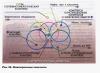
Lightning teeth - nucleotides (Adenine-Thymine/Uracil/, Guanine-Cytazine). All lightning is DNA.

To transfer information from DNA, 2 strands must be broken. The bond between A-T and G-C is hydrogen, therefore it is easily broken by the enzyme Helicase:

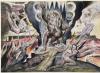
To prevent knots from forming (I twisted a towel as an example):


To prevent the chain from twisting, one DNA strand at the origin of replication is cut by Topoisomerase.

When one thread is free, the second can easily rotate around its axis, thereby relieving tension during “unwinding”. Nodes appear, energy is saved.
Then, an RNA primer is needed to start assembling the RNA. The protein that assembles mRNA cannot simply assemble the first nucleotide, it needs a piece of RNA to start (it’s written there in detail, I’ll write it out later). This piece is called the RNA primer. And this protein already attaches the first nucleotide to it.