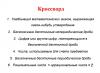
It's no secret that athletes, even in good physical fitness and comparatively early age often quit training due to injuries. Most of their problems are ligaments. Their weakest part is cartilage tissue. It turns out that the functions of damaged joints can be restored if you pay attention to the problem in time and create suitable conditions for the treatment and regeneration of their cells.
Tissues in the human body
The human body is a complex and flexible system capable of self-regulation. It consists of cells of different structure and functions. The main metabolism takes place in them. Together with non-cellular structures they are combined into tissues: epithelial, muscle, nervous, connective.
Epithelial cells form the basis of the skin. They line the internal cavities (abdominal, thoracic, upper respiratory tract, intestinal tract). Muscle tissue allows a person to move. It also ensures the movement of internal media in all organs and systems. Musculature is divided into types: smooth (walls of abdominal organs and blood vessels), cardiac, skeletal (striated). Nervous tissue ensures the transmission of impulses from the brain. Some cells are capable of growing and multiplying, some of them are capable of regeneration.
Connective tissue is the internal environment of the body. It is different in structure, structure and properties. It consists of strong skeletal bones, subcutaneous fatty tissue, liquid media: blood and lymph. It also includes cartilage tissue. Its functions are formative, shock-absorbing, supporting and supporting. They all play important role and are necessary in complex system body.
structure and functions
Her characteristic feature- looseness in the arrangement of cells. Looking at them separately, you can see how clearly they are separated from each other. The ligament between them is the intercellular substance - the matrix. Moreover, different types it is formed of cartilage in addition to the main one amorphous substance various fibers (elastic and collagen). Although they have a common protein origin, they differ in properties and, depending on this, perform different functions.

All bones in the body are formed from cartilage. But as they grew, their intercellular substance became filled with salt crystals (mainly calcium). As a result, the bones gained strength and became part of the skeleton. Cartilage also performs supporting functions. In the spine, being between the segments, they perceive constant loads (static and dynamic). The ears, nose, trachea, bronchi - in these areas the tissue plays a more formative role.
The growth and nutrition of cartilage occurs through the perichondrium. She is in the fabric mandatory part except for the joints. They contain synovial fluid between the rubbing surfaces. It washes, lubricates and nourishes them, removes waste products.
Structure
In cartilage there are few cells capable of dividing, and there is a lot of space around them, filled with protein substances of varying properties. Due to this feature, regeneration processes are often to a greater extent go exactly in the matrix.
There are two types of tissue cells: chondroblasts (mature) and chondroblasts (young). They differ in size, location and location. Chondrocytes have a round shape and are larger. They are located in pairs or in groups of up to 10 cells. Chondroblasts are usually smaller and are found peripherally or singly in the tissue.
Water accumulates in the cytoplasm of cells under the membrane, and there are inclusions of glycogen. Oxygen and nutrients enter the cells diffusely. There the synthesis of collagen and elastin occurs. They are necessary for the formation of intercellular substance. It depends on its specificity what type of cartilage tissue it will be. The structural features differ from intervertebral discs, including the collagen content. In the nasal cartilage, the intercellular substance consists of 30% elastin.

Species
How it is classified Its functions depend on the predominance of specific fibers in the matrix. If there is more elastin in the intercellular substance, then the cartilage tissue will be more plastic. It is almost as strong, but the fiber bundles in it are thinner. They withstand loads not only in compression, but also in tension, and are capable of deformation without critical consequences. Such cartilages are called elastic. Their tissues form the larynx, ears, and nose.
If in the matrix around the cells great content collagen with complex structure building polypeptide chains, such cartilage is called hyaline. It most often covers internal surfaces joints. Largest quantity collagen is concentrated in the superficial zone. It plays the role of a frame. The structure of the fiber bundles in it resembles three-dimensional intertwined networks of a spiral shape.
There is another group: fibrous, or fibrous, cartilage. They, like hyaline ones, contain a large amount of collagen in the intercellular substance, but it has a special structure. The bundles of their fibers do not have a complex weave and are located along the axis of the greatest loads. They are thicker, have special compressive strength, and do not recover well when deformed. Intervertebral discs, the junction of tendons and bones, are formed from such tissue.

Functions
Due to its special biomechanical properties, cartilage tissue is ideal for connecting the components of the musculoskeletal system. It is capable of receiving the effects of compression and tension forces during movements, redistributing them evenly to the load, and to some extent absorbing or dissipating them.
Cartilage forms abrasion-resistant surfaces. Together with synovial fluid, such joints, under acceptable loads, are able to perform their functions normally for a long time.
Tendons are not cartilage tissue. Their functions also include linking into a common apparatus. They also consist of bundles of collagen fibers, but their structure and origin are different. respiratory organs, ears, in addition to performing formative and supporting functions, are a place of attachment of soft tissues. But unlike tendons, the muscles next to them do not have the same load.
Special properties
Elastic cartilage has very few blood vessels. And this is understandable, because a strong dynamic load can damage them. How is cartilage connective tissue nourished? These functions are performed by the intercellular substance. There are no vessels at all in hyaline cartilage. Their rubbing surfaces are quite hard and dense. They are nourished by the synovial fluid of the joint.
Water moves freely in the matrix. It contains all the necessary substances for metabolic processes. Proteoglycan components in cartilage perfectly bind water. As an incompressible substance, it provides rigidity and additional shock absorption. When under load, water takes on the impact, spreads throughout the intercellular space and smoothly relieves stress, preventing irreversible critical deformations.

Development
In the body of an adult, up to 2% of the mass is cartilage tissue. Where is it located and what functions does it perform? Cartilage and bone tissue do not differentiate in the embryonic period. Fetuses have no bones. They develop from cartilage tissue and are formed at the time of birth. But part of her never ossifies. From it the ears, nose, larynx, and bronchi are formed. It is also present in the joints of the arms and legs, articulations of the intervertebral discs, and menisci of the knees.
The development of cartilage occurs in several stages. First, mesenchymal cells become saturated with water, become rounded, lose their processes, and begin to produce substances for the matrix. After this, they differentiate into chondrocytes and chondroblasts. The former are tightly surrounded by intercellular substance. In this state they can divide limited quantity once. After such processes, an isogenic group is formed. The cells remaining on the surface of the tissue become chondroblasts. In the process of producing matrix substances, final differentiation occurs, a structure is formed with a clear division into a thin border and the base of the tissue.

Age-related changes
The functions of cartilage do not change during life. However, over time, you can notice signs of aging: the muscles and tendons of the joints weaken, flexibility is lost, and pain occurs when the weather changes or during unusual exercise. This process is considered a physiological norm. By the age of 30-40 years, the symptoms of changes may already begin to cause inconvenience to a greater or lesser extent. Aging of articular cartilage tissue occurs due to loss of its elasticity. The elasticity of the fibers is lost. The fabric dries and becomes loose.
Cracks appear on the smooth surface and it becomes rough. Smoothness and ease of gliding is no longer possible. The damaged edges grow, deposits form in them, and osteophytes form in the tissue. Elastic cartilages age with the accumulation of calcium in the intercellular substance, but this has almost no effect on their functions (nose, ears).

Dysfunction of cartilage and bone tissue
When and how can this happen? IN to a large extent it depends on what function the cartilage tissue performs. In intervertebral discs, the main function of which is stabilizing and supporting, disruption most often occurs with the development of dystrophic or degenerative processes. The situation can lead to displacements, which, in turn, will lead to compression of surrounding tissues. Swelling, pinched nerves, and compression of blood vessels are inevitable.
To restore stability, the body tries to fight the problem. The vertebra at the site of deformation “adjusts” to the situation and grows in the form of peculiar bone outgrowths (whiskers). This also does not benefit the surrounding tissues: again swelling, pinching, compression. This problem is complex. Disturbances in the functioning of the osteochondrosis apparatus are commonly called osteochondrosis.
Long-term restriction of movement (plaster for injuries) also negatively affects cartilage. If, under excessive loads, elastic fibers degenerate into coarse fibrous bundles, then with low activity, the cartilage stops feeding normally. Synovial fluid does not mix well, chondrocytes do not receive enough nutrients, and as a result they are not produced required quantity collagen and elastin for the matrix.
The conclusion suggests itself: for normal joint function, cartilage must receive sufficient tension and compression load. To ensure this, you need to engage physical exercise, lead a healthy and active lifestyle.
CARTILAGE TISSUE
General characteristics: relatively low level metabolism, absence of blood vessels, hydrophilicity, strength and elasticity.
Structure: chondrocyte cells and intercellular substance (fibers, amorphous substance, interstitial water).
Lecture: CARTILAGE TISSUE
Cells ( chondrocytes) constitute no more than 10% of the cartilage mass. The main volume in cartilage tissue is accounted for intercellular substance. The amorphous substance is quite hydrophilic, which allows it to deliver nutrients to cells by diffusion from the capillaries of the perichondrium.
Chondrocyte differon: stem, semi-stem cells, chondroblasts, young chondrocytes, mature chondrocytes.
Chondrocytes are derivatives of chondroblasts and the only population of cells in cartilage tissue, located in the lacunae. Chondrocytes can be divided according to their maturity into young and mature. Young retain the structural features of chondroblasts. They have an oblong shape, a developed GREPS, a large Golgi apparatus, and are capable of forming proteins for collagen and elastic fibers and sulfated glycosaminoglycans and glycoproteins. Mature chondrocytes have an oval or round shape. The synthetic apparatus is less developed when compared to young chondrocytes. Glycogen and lipids accumulate in the cytoplasm.
Chondrocytes are capable of dividing and form isogenic groups of cells surrounded by a single capsule. In hyaline cartilage, isogenic groups can contain up to 12 cells, in elastic and fibrous cartilage - smaller number cells.
Functions cartilaginous tissues: supporting, formation and functioning of joints.
Classification of cartilage tissues
There are: 1) hyaline, 2) elastic and 3) fibrous cartilaginous tissue.
Histogenesis . During embryogenesis, cartilage is formed from mesenchyme.
1st stage. Formation of a chondrogenic island.
2nd stage. Differentiation of chondroblasts and the beginning of the formation of fibers and cartilage matrix.
3rd stage. The growth of cartilage anlage in two ways:
1) Interstitial growth– caused by an increase in tissue from the inside (formation of isogenic groups, accumulation of the intercellular matrix), occurs during regeneration and in the embryonic period.
2) Appositional growth– caused by tissue layering due to the activity of chondroblasts in the perichondrium.
Cartilage regeneration . When cartilage is damaged, regeneration occurs from the cambial cells in the perichondrium, and new layers of cartilage are formed. Complete regeneration occurs only in childhood. Adults are characterized by incomplete regeneration: PVNST is formed in place of the cartilage.
Age-related changes . Elastic and fibrous cartilage are resistant to damage and change little with age. Hyaline cartilage tissue can undergo calcification, sometimes transforming into bone tissue.
Cartilage as an organ consists of several tissues: 1) cartilage tissue, 2) perichondrium: 2a) outer layer - PVST, 2b) inner layer - PBST, with blood vessels and nerves, and also contains stem, semi-stem cells and chondroblasts.
1. HYALINE CARTILAGE TISSUE
Localization: cartilages of the nose, larynx (thyroid cartilage, cricoid cartilage, arytenoid, except vocal processes), trachea and bronchi; articular and costal cartilages, cartilaginous growth plates in tubular bones.
Structure: cartilage cells, chondrocytes (described above) and intercellular substance, consisting of collagen fibers, proteoglycans and interstitial water. Collagen fibers(20-25%) consist of type II collagen and are arranged randomly. Proteoglycans, constituting 5-10% of the mass of cartilage, they are represented by sulfated glycosaminoglycans, glycoproteins that bind water and fiber. Proteoglycans of hyaline cartilage prevent its mineralization. Interstitial water(65-85%) ensures the incompressibility of cartilage and acts as a shock absorber. Water promotes efficient metabolism in cartilage, transports salts, nutrients, and metabolites.
Articular cartilage is a type of hyaline cartilage, does not have perichondrium, and receives nutrition from synovial fluid. In articular cartilage there are: 1) a superficial zone, which can be called acellular, 2) a middle (intermediate) zone - containing columns cartilage cells and 3) the deep zone where cartilage interacts with bone.
I suggest watching the video from YouTube " ARTHROSIS OF THE KNEE JOINT»
2. ELASTIC CARTILAGE TISSUE
Localization: auricle, cartilages of the larynx (epiglottic, corniculate, sphenoid, as well as the vocal process at each arytenoid cartilage), eustachian tube. This type of tissue is necessary for those areas of organs that are capable of changing their volume, shape and have reversible deformation.
Structure: cartilage cells, chondrocytes (described above) and intercellular substance, consisting of elastic fibers (up to 95%) fibers and amorphous substance. For imaging, dyes that reveal elastic fibers, such as orcein, are used.
3. FIBROUS CARTILAGE TISSUE
Localization: fibrous rings of intervertebral discs, articular discs and menisci, in the symphysis (symphysis pubis), articular surfaces in the temporomandibular and sternoclavicular joints, in places of attachment of tendons to bones or hyaline cartilage.
Structure: chondrocytes (usually singly) of an elongated shape and intercellular substance, consisting of a small amount of amorphous substance and a large number of collagen fibers. The fibers are arranged in orderly parallel bundles.
70.Cartilage tissue. Classification, development, structure, histochemical characteristics and function. Cartilage growth, regeneration and age-related changes.
Cartilaginous And bone tissue develop from sclerotomal mesenchyme, belong to tissues internal environment and, like all other tissues of the internal environment, consist of cells and intercellular substance. The intercellular substance here is dense, so these tissues perform a support-mechanical function.
Cartilage tissue(textuscartilagineus). They are classified into hyaline, elastic and fibrous. The classification is based on the peculiarities of the organization of the intercellular substance. The composition of cartilage tissue includes 80% water, 10-15% organic substances and 5-7% inorganic substances.
Development of cartilage tissue, or chondrogenesis, consists of 3 stages: 1) formation of chondrogenic islets; 2) formation of primary cartilaginous tissue: 3) differentiation of cartilaginous tissue.
During 1st stage mesenchymal cells unite into chondrogenic islands, the cells of which multiply and differentiate into chondroblasts. The resulting chondroblasts contain granular ER, Golgi complex, and mitochondria. Chondroblasts then differentiate into chondrocytes.
During 2nd stage In chondrocytes, granular ER, Golgi complex, and mitochondria are well developed. Chondrocytes actively synthesize fibrillar protein (type II collagen), from which the intercellular substance is formed, which stains oxyphilic.
When advancing 3rd stage in chondrocytes, granular ER develops more intensively, on which fibrillar proteins and chondroitin sulfates (chondroitinsulfuric acid) are produced, which are stained with basic dyes. Therefore, the main intercellular substance of the cartilage tissue around these chondrocytes is stained basophilic.
Around the cartilaginous rudiment, a perichondrium is formed from mesenchymal cells, consisting of 2 layers: 1) the outer, more dense, or fibrous, and 2) the inner, more loose, or chondrogenic, which contains prechondroblasts and chondroblasts.
Appositional growth of cartilage, or growth by superposition, is characterized by the fact that chondroblasts are released from the perichondrium, which superimpose on the main substance of the cartilage, differentiate into chondrocytes and begin to produce the intercellular substance of cartilage tissue.
Interstitial growth cartilage tissue is produced by chondrocytes located inside the cartilage, which, firstly, divide by mitosis and, secondly, produce intercellular substance, due to which the volume of cartilage tissue increases.
Cartilage cells(chondrocytus). The differentiaten of chondrocytes is composed of: stem cell, semi-stem cell (prechondroblast), chondroblast, chondrocyte.
Chondroblasts (chondroblastus) are located in the inner layer of the perichondrium and have organelles of general importance: granular ER, Golgi complex, mitochondria. Functions of chondroblasts:
1) secrete intercellular substance (fibrillar proteins);
2) in the process of differentiation they turn into chondrocytes;
3) have the ability to undergo mitotic division.
Chondrocytes located in cartilaginous lacunae. In the lacuna there is initially 1 chondrocyte, then, during its mitotic division, 2, 4, 6, etc. cells are formed. All of them are located in the same lacuna and form an isogenic group of chondrocytes.
Chondrocytes of the isogenic group are divided into 3 types: I, II, III.
Type I chondrocytes have the ability to undergo mitotic division, contain the Golgi complex, mitochondria, granular ER and free ribosomes, have a large nucleus and small quantity cytoplasm (large nuclear-cytoplasmic ratio). These chondrocytes are located in young cartilage.
Type II chondrocytes located in mature cartilage, their nuclear-cytoplasmic ratio decreases somewhat as the volume of the cytoplasm increases; they lose the ability to undergo mitosis. Granular EPS is well developed in their cytoplasm; they secrete proteins and glycosaminoglycans (chondroitin sulfates), so the main intercellular substance around them is stained basophilic.
Chondrocytes III type are located in old cartilage, lose the ability to synthesize glycosaminoglycans and produce only proteins, therefore the intercellular substance around them is stained oxyphilic. Consequently, around such an isogenic group one can see an oxyphilic-stained ring (proteins are secreted by type III chondrocytes), outside of this ring a basophilic-stained ring is visible (glycosaminoglycans are secreted by type II chondrocytes) and the outer ring itself is again oxyphilic-stained (proteins are secreted at a time when cartilage contained only young type I chondrocytes). Thus, these 3 differently colored rings around isogenic groups characterize the process of formation and function of 3 types of chondrocytes.
Intercellular substance of cartilage tissue. Contains organic matter(mainly type II collagen), glycosaminoglycans, proteoglycans and non-collagen type proteins. The more proteoglycans, the more hydrophilic the intercellular substance, the more elastic and permeable it is. Gases, water molecules, salt ions and micromolecules diffusely penetrate through the ground substance from the side of the perichondrium. However, macromolecules do not penetrate. Macromolecules have antigenic properties, but since they do not penetrate the cartilage, cartilage transplanted from one person to another takes root well (no immune rejection reaction occurs).
The main substance of cartilage contains collagen fibers consisting of type II collagen. The orientation of these fibers depends on power lines, and the direction of the latter depends on mechanical impact on cartilage In the intercellular substance of cartilage tissue there are no blood and lymphatic vessels, therefore the nutrition of the cartilage tissue is carried out through the diffuse supply of substances from the vessels of the perichondrium.
Age-related changes in cartilage tissue. The greatest changes are observed in old age, when the number of chondroblasts in the perichondrium and the number of dividing cartilage cells decrease. In chondrocytes, the amount of granular ER, Golgi complex and mitochondria decreases, and the ability of chondrocytes to synthesize glycosaminoglycans and proteoglycans is lost. A decrease in the amount of proteoglycans leads to a decrease in the hydrophilicity of cartilage tissue, a weakening of cartilage permeability and nutrients. This leads to calcification of the cartilage, penetration of blood vessels into it and the formation of bone substance inside the cartilage.
Hello my friends!
In this article we will look at what it is knee joint cartilage. Let's look at what cartilage is made of and what its function is. As you understand, in all joints of our body the cartilage tissue is the same, and everything described below also applies to other joints.
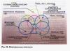
The ends of our bones in the knee joint are covered with cartilage, between them lie two menisci - these are also cartilages, but only slightly different in composition. Read about menisci in the article "". I will only say that cartilage and menisci differ in the type of cartilage tissue: bone cartilage is hyaline cartilage, and the menisci – fibrocartilage. This is what we will look at now.
The thickness of the cartilage covering the ends of the bone is on average 5-6 mm, it consists of several layers. Cartilage is dense and smooth, which allows bones to easily slide against each other during flexion and extension movements. Possessing elasticity, cartilage acts as a shock absorber during movements.
In a healthy joint, depending on its size, the fluid content is from 0.1 to 4 ml, the distance between the cartilages (articular space) is from 1.5 to 8 mm, acid-base balance 7.2-7.4, water 95%, protein 3%. The composition of cartilage is similar to blood serum: 200-400 leukocytes per 1 ml, of which 75% are lymphocytes.
Cartilage is one of the types of connective tissue in our body. The main difference between cartilage tissue and others is the absence of nerves and blood vessels that directly feed this tissue. Blood vessels would not withstand the stress and constant pressure, and the presence of nerves there would be reflected in pain with every movement we made.
Cartilage is designed to reduce friction where bones connect. Cover both heads of the bone and inner side patella (patella). Constantly washed by synovial fluid, they ideally reduce friction in the joints to zero.
Cartilage does not have access to blood vessels and nutrition, respectively, and if there is no nutrition, then there is no growth or repair. But cartilage also consists of living cells and they also need nutrition. They receive nutrition from the same synovial fluid.
The meniscus cartilage is riddled with fibers, which is why it is called fibrocartilage and is denser and harder in structure than hyaline, therefore it has greater tensile strength and can withstand pressure.
Cartilage differs in its fiber ratio: . All this gives the cartilage not so much hardness as elasticity. Working like a sponge under load, cartilage and menisci are compressed, unclenched, flattened, stretched as you wish. They constantly absorb a new portion of liquid and give away the old one, forcing it to constantly circulate; at the same time, the liquid is enriched with nutrients and again carries them to the cartilage. We'll talk about synovial fluid later.
Main components of cartilage
Articular cartilage - This is a complex fabric in its structure. Let's look at the main components of this fabric. make up almost half of the intercellular space in articular cartilage. Collagen in its structure consists of very large molecules, intertwined in triple spirals. This structure of collagen fibers allows cartilage to resist any type of deformation. Collagen gives tissue elasticity. give elasticity, the ability to return to its original state.
Second having great importance cartilage element – water, which is in large quantities contained in intercellular space. Water is unique natural element, it is not subject to any deformation, it can neither be stretched nor compressed. This adds rigidity and elasticity to the cartilage tissue. Moreover, than more water, the better and more functional the interarticular fluid. It spreads and circulates easily. With a lack of water, the joint fluid becomes more viscous, less fluid and, of course, performs its role in providing nutrition to the cartilage worse. !
Glycosamines– substances produced by the cartilage tissue of the joints are also part of the synovial fluid. By its structure, glucosamine is a polysaccharide and serves as an important component of cartilage.
Glucosamine is a precursor of glycosaminoglycans (the main component of articular cartilage), so it is believed that its additional use from the outside can contribute to the restoration of cartilage tissue.
In our body, glucosamine binds cells and is part of cell membranes and proteins, making fabrics stronger and more resistant to stretching. Thus, glucosamine supports and strengthens our joints and ligaments. With a decrease in the amount of glucosamines, the resistance of cartilage tissue to stress also decreases, and the cartilage becomes more sensitive to damage.
The issues of restoration of cartilage tissue and the production of necessary compounds and substances are dealt with chondrocytes.
Chondrocytes, by their nature, do not differ from other cells in terms of development and regeneration, their metabolic rate is quite high. But the problem is that there are very few of these same chondrocytes. In articular cartilage, the number of chondrocytes is only 2-3% of the mass of the cartilage. Therefore, the restoration of cartilage tissue is so limited.
So, nutrition of cartilage is difficult, renewal of cartilage tissue is also a very long-term process, and restoration is even more problematic. What to do?
Considering all of the above, we come to the conclusion that in order for the knee joint cartilage to recover, it is necessary to achieve high numbers and activity of chondrocyte cells. And our task is to provide them with adequate nutrition, which they can only receive through synovial fluid. But, even if the nutrition is the richest, it will not achieve its goal without moving the joint. That's why, If you move more, your recovery will be better!
With prolonged immobilization of a joint or the entire leg (plaster, splints, etc.), not only the muscles decrease and atrophy; It has been established that cartilage tissue also decreases, since it does not receive enough nutrition without movement. I will repeat myself for the hundredth time, but this is yet another proof of the need for constant movement. Man is created by nature in such a way that he must constantly run for food and run away from the mammoth, like other animals. Excuse me if I offend some of the “Crowns of Nature” by this. To scale evolutionary development, we have come too far for the body to behave differently; it has not yet adapted to other conditions of existence. And if the body feels that something in its composition is not needed or does not work well, it gets rid of it. Why feed something that is not beneficial? They stopped walking with their legs - their legs atrophied, the bodybuilder stopped pumping (using all his muscle mass) - he immediately deflated. Well, I got distracted.
In other articles, we will, of course, touch on issues (surgical and conservative methods), their nutrition and movement. This is what I, with my cartilage injury, am trying to implement. I'll tell you too.
Well, for now my instructions: , COMPLETE VARIED NUTRITION,.
You can start right now.
All the best, don't get sick!
Cartilaginous and bone tissues develop from sclerotomal mesenchyme; they belong to the tissues of the internal environment and, like all tissues of the internal environment, consist of cells and intercellular substance. The intercellular substance here is dense, so these tissues perform a support-mechanical function.
CARTILAGE TISSUE (textus cartilagineus) is classified into hyaline, elastic and fibrous. The classification is based on the peculiarities of the organization of intercellular substance. The composition of cartilage tissue includes 80% water, 10-15% organic substances and 5-7% inorganic substances.
DEVELOPMENT OF CARTILAGE TISSUE or CHONDROGENESIS consists of 3 stages:
ü formation of chondrogenic islets;
ü formation of primary cartilage tissue;
ü differentiation of cartilage tissue.
During STAGE 1, mesenchymal cells unite into chondrogenic islands, the cells of which multiply and differentiate into chondroblasts. The resulting chondroblasts contain granular ER, Golgi complex, and mitochondria. Chondroblasts then differentiate into chondrocytes.
2nd STAGE. In chondrocytes, granular ER, Golgi complex, and mitochondria are well developed. Chondrocytes actively synthesize fibrillar protein (type I collagen), from which the intercellular substance is formed, which stains oxyphilic.
With the onset of STAGE 3, granular EPS develops more intensively in chondrocytes, which produces both fibrillar proteins and chondriatine sulfates (chondriatin sulfuric acid), which are stained with basic dyes. Therefore, the main intercellular substance of the cartilage tissue around these chondrocytes is stained basophilic.
Around the cartilaginous rudiment, a perichondrium is formed from mesenchymal cells, consisting of 2 layers: 1) the outer, more dense, or fibrous, and 2) the inner, more loose, or chondrogenic, which contains prechondroblasts and chondroblasts.
APPOSITIONAL GROWTH OF CARTILAGE, or growth by superposition, is characterized by the fact that chondroblasts are released from the perichondrium, which superimpose on the main substance of the cartilage, differentiate into chondrocytes and begin to produce the intercellular substance of cartilage tissue.
INTERSTITIAL GROWTH cartilage tissue is produced by chondrocytes located inside the cartilage, which, firstly, divide by mitosis and, secondly, produce intercellular substance, due to which the volume of cartilage tissue increases.
CARTILAGE TISSUE CELLS(chondrocytus) constitute the differential of chondrocytes: stem cell, semi-stem cell (prechondroblast), chondroblast, chondrocyte.
CHONDROBLASTS(chondroblastocytus) are in inner layer perichondrium, have organelles general meaning: granular ER, Golgi complex, mitochondria. FUNCTION of chondroblasts: 1) secrete intercellular substance (fibrillar proteins); 2) in the process of differentiation they turn into chondrocytes; 3) have the ability to undergo mitotic division.
CHONDROCYTES located in cartilaginous lacunae. In the lacuna there is initially 1 chondrocyte, then during its mitotic division 2, 4, 6, etc. are formed. cells. All of them are located in the same lacuna and form an isogenic group of chondrocytes.
Chondrocytes of the isogenic group are divided into 3 types: I, II, III.
CHONDROCYTES TYPE I have the ability to undergo mitotic division, contain the Golgi complex, mitochondria, granular EPS and free ribosomes, have a large nucleus and a small amount of cytoplasm (large nuclear-cytoplasmic ratio). These chondrocytes are located in young cartilage.
TYPE II CHONDROCYTES are located in mature cartilage, their nuclear-cytoplasmic ratio decreases somewhat, as the volume of the cytoplasm increases, they lose the ability to mitosis. Granular EPS is well developed in their cytoplasm; they secrete proteins and glycosaminoglycans (chondriatin sulfates). Therefore, the main intercellular substance around them is stained basophilic.
TYPE III CHONDROCYTES are located in old cartilage, lose the ability to synthesize glycosaminoglycans and produce only proteins, therefore the intercellular substance around them is stained oxyphilic. Consequently, around such an isogenic group one can see an oxyphilic-stained ring (proteins are secreted by type 3 chondrocytes, a basophilic-stained ring is visible outside this ring), (glycosaminoglycans are secreted by type 2 chondrocytes) and the outermost ring is again oxyphilic-stained (proteins are secreted while there were only young type 1 chondrocytes in the cartilage). Thus, these 3 differently colored rings around isogenic groups characterize the process of formation and function of 3 types of chondrocytes.
INTERCELLULAR SUBSTANCE OF CARTILAGE TISSUE contains organic substances (mainly type II collagen), glycosaminoglycans, proteoglycans and non-collagen type proteins. The more proteoglycans, the more hydrophilic the intercellular substance, the more elastic it is and the more permeable it is. Gases, water molecules, salt ions and micromolecules diffusely penetrate through the ground substance from the side of the perichondrium. However, macromolecules do not penetrate. Macromolecules have antigenic properties. But since they do not penetrate the cartilage, cartilage transplanted from one person to another takes root well (an immune rejection reaction does not occur).
The main substance of cartilage contains collagen fibers consisting of type II collagen. The orientation of these fibers depends on the force lines, and the direction of the force lines depends on the mechanical force on the cartilage. In the intercellular substance of cartilage tissue there are no blood and lymphatic vessels, therefore the nutrition of the cartilage tissue is carried out through the diffuse supply of substances from the vessels of the perichondrium.
HYALINE CARTILAGE TISSUE has a bluish-whitish color, translucent, fragile, in the body it is found at the junction of the ribs with the sternum, in the walls of the trachea and bronchi, larynx, and on the articular surfaces. Depending on where the hyaline cartilage is located, it has different structure. If there is a malnutrition, the hyaline cartilage undergoes calcification.
HYALINE CARTILAGE AT THE ENDS OF THE RIBS covered with perichondrium, under which there is a zone of young cartilage. Here are young spindle-shaped chondrocytes located in cartilaginous lacunae and capable of producing only fibrillar proteins. Therefore, the intercellular substance around them is oxyphilic colored. The deeper chondrocytes become rounder. Even deeper, isogenic groups of chondrocytes are formed, capable of producing proteins and chondriatic sulfuric acid, which stains basophilically. Therefore, the intercellular substance around them is stained with basic dyes. Even deeper are isogenic groups containing even more mature chondrocytes that secrete only proteins. Therefore, the ground substance around them is colored oxyphilic.
HYALINE CARTILAGE OF ARTICULAR SURFACES does not have perichondrium and consists of 3 zones that are not clearly demarcated from each other. The outer zone includes spindle-shaped chondrocytes located in lacunae parallel to the surface of the cartilage. Deeper is the columnar zone, the cells of which continuously divide and form columns and inner zone divided by the basophilic line into non-calcified and calcified parts. Calcified part adjacent to bone tissue, contains matrix vesicles and blood vessels.
NUTRITION This cartilage is formed from 2 sources: 1) due to nutrients found in the synovial fluid of the joint and 2) due to blood vessels passing through the calcified cartilage.
ELASTIC CARTILAGE TISSUE has a whitish-yellowish color, located in auricle, the wall of the external auditory canal, the arytenoid and corniculate cartilages of the larynx, the epiglottis, and in the medium-caliber bronchi. It differs from hyaline cartilage in that, firstly, it is elastic, since in addition to collagen, it contains elastic fibers that go into various directions and interwoven into the perichondrium, and stained with orcein in brown; secondly, it contains less chondriatic sulfuric acid, lipids and glycogen; thirdly, it never undergoes calcification. At the same time general plan The structure of elastic cartilage tissue is similar to hyaline cartilage.
FIBER CARTILAGE(cortilago fibrosa) is located in the intervertebral discs, pubic fusion, sites of attachment of tendons to hyaline cartilage and in the maxillary joints. This cartilage is characterized by the presence of 3 sections: 1) tendon part; 2) fibrocartilage itself; 3) hyaline cartilage. Where there is a tendon, bundles of collagen fibers run parallel to each other, fibrocytes are located between them; in fibrous cartilage tissue the parallel arrangement of fibers is maintained, chondrocytes are located in the lacunae of the cartilaginous substance; hyaline cartilage has a normal structure.
AGE CHANGES IN CARTILAGE TISSUE. Biggest changes observed in old age, when the number of chondroblasts in the perichondrium and the number of dividing cartilage cells decrease. In chondrocytes, the amount of granular ER, Golgi complex and mitochondria decreases, and the ability of chondrocytes to synthesize glycosaminoglycans and proteoglycans is lost. A decrease in the amount of proteoglycans leads to a decrease in the hydrophilicity of cartilage tissue, weakening the permeability of cartilage and the supply of nutrients. This leads to calcification of the cartilage, penetration of blood vessels into it and the formation of bone substance inside the cartilage.