However, we must also keep in mind that, like other research methods, x-ray diagnostics has its own capabilities and disadvantages. Along with the x-ray picture, characteristic of a particular pathological process or even pathognomonistic, during the study there is almost the same x-ray image with various diseases. For example, a lung tumor, enlargement of bifurcation lymph nodes and blockage in the thoracic part of the esophagus, when they coincide in place with the bifurcation area on the screen or x-ray, are difficult to differentiate. The same thing happens with pneumonia and diaphragmatic hernia, if you do not see the patient and do not examine him clinically.
Therefore, any X-ray examination should always be preceded by a careful collection of anamnestic data and a comprehensive thorough clinical trial. The final diagnosis must always be made by comparing data from all research methods.
Based on all this, X-ray examination, as very important method, should neither be underestimated nor overestimated.
This section of this book concerns a number of general issues X-ray diagnostics, characterizing the methods and capabilities of X-ray examinations, as well as low-power X-ray machines suitable for examining dogs.
Nature x-rays
Rays, which are now called X-rays, were discovered on November 7, 1895 by physicist V. K. Roentgen. The official date of discovery of these rays is December 28, 1895, when Roentgen, after studying the X-rays he discovered, published the first report on their properties.
These X-rays began to be called X-rays on January 23, 1896, when V. K. Roentgen made a public report on X-rays at a meeting of the Physico-Medical Society. At this meeting, it was unanimously decided to call X-rays X-rays.
The nature of X-rays remained little studied for 17 years from the date of their discovery by V.K. Roentgen, although soon after the discovery of these rays the scientist himself and a number of other researchers noted their similarity with visible rays.
The similarity was confirmed by the straightness of propagation, the absence of deviations in electrical and magnetic fields. But, on the other hand, it was not possible to detect either the phenomenon of refraction by a prism, or reflection from mirrors and a number of other properties characteristic of visible light, which has a wave nature.
And only in 1912, first our compatriot, the famous Russian physicist A.I. Lebedev, and then the German physicist Laue, managed to prove that X-rays are of the same nature as rays of visible light, that is, they are electromagnetic waves. Thus, X-rays are the same in nature as radio waves, infrared rays, visible light rays and ultraviolet rays.
The only difference between these rays is that they have different wavelengths electromagnetic vibrations. Among the above, X-rays have a very short wavelength. That's why they demanded special conditions producing experiments to identify refraction or reflection.
The wavelength of X-rays is measured in a very small unit called an angstrom (1 Å = 10–8 cm, that is, one hundred millionth of a centimeter). In practice, diagnostic devices produce rays with a wavelength of 0.1–0.8 Å.
Properties of X-rays
X-rays pass through opaque bodies and objects, such as, for example, paper, matter, wood, tissues of the human and animal body, and even metals of a certain thickness. Moreover, the shorter the wavelength of radiation, the easier they pass through the listed bodies and objects.
In turn, when these rays pass through bodies and objects with different densities, they are partially absorbed. Dense bodies absorb X-rays more intensely than low-density bodies.
X-rays have the ability to excite visible glow some chemical substances. For example: barium platinum cyanide crystals, when exposed to X-rays, begin to glow with a bright greenish-yellowish light. The glow continues only at the moment of exposure to x-rays and immediately stops with the cessation of irradiation. Platinum cyanide barium thus fluoresces from the action of X-rays. (This phenomenon was the reason for the discovery of X-rays.)
When illuminated with x-rays, calcium tungstic acid also glows, but with blue light, and the glow of this salt continues for some time even after the irradiation has stopped, i.e. phosphorescent.
The property of causing fluorescence is used to produce transillumination using X-rays. The ability to cause phosphorescence in some substances is used to produce x-rays.
X-rays also have the ability to act on the photosensitive layer of photographic plates and films like visible light, causing the decomposition of silver bromide. In other words, these rays have a photo-chemical effect. This circumstance makes it possible to take photographs using X-rays from various parts of the body in humans and animals.
X-rays have a biological effect on the body. Passing through a certain area of the body, they produce corresponding changes in tissues and cells depending on the type of tissue and the amount of rays absorbed by them, i.e. the dose.
This property is used to treat a number of diseases in humans and animals. When exposed to large doses of X-rays in the body, a whole range of functional and morphological changes, and a specific disease arises - radiation sickness.
X-rays, in addition, have the ability to ionize air, that is, split the constituent parts of air into separate, electrically charged particles.
As a result, the air becomes an electrical conductor. This property is used to determine the number of X-rays emitted by an X-ray tube per unit of time using special devices - dosimeters.
Knowing the radiation dose from the X-ray tube is important when X-ray therapy is performed. Without knowing the radiation dose of the tube at the appropriate rigidity, it is impossible to carry out treatment with X-rays, since instead of improving, it is easy to worsen the entire disease process. Improper use of X-rays for treatment can destroy healthy tissue and even cause serious damage throughout the body.
Methods of X-ray examination
A) Transillumination (fluoroscopy). X-rays are used in veterinary practice to study and recognize various diseases in farm animals. This method of studying sick animals is auxiliary to establish or clarify the diagnosis along with other methods. Therefore the data x-ray examination always needs to be linked to data from clinical and other studies. Only in this case can we come to correct conclusion and accurate diagnosis. As stated above, there are two methods of x-ray examination: the first method is transillumination or fluoroscopy, the second method is the production of x-rays or radiography.
Let us dwell on the issue of justification for transillumination, the possibilities of this method, its advantages and disadvantages.
In order to perform transillumination with invisible X-rays and obtain a visible shadow picture of the examined area of the body, they use certain properties X-rays and body tissues.
1. The ability of X-rays: a) to penetrate body tissues, and b) to cause visible luminescence of certain chemical substances.
2. The ability of tissues to absorb x-rays to one degree or another depending on their density.
Radiology is a branch of radiology that studies the effects of x-ray radiation on the body of animals and humans resulting from this disease, their treatment and prevention, as well as methods for diagnosing various pathologies using x-rays (x-ray diagnostics). A typical X-ray diagnostic apparatus includes a power supply (transformers), a high-voltage rectifier, a converter alternating current electrical network in a permanent one, control panel, tripod and x-ray tube.
X-rays are a type of electromagnetic oscillations that are formed in an X-ray tube during a sharp deceleration of accelerated electrons at the moment of their collision with atoms of the anode substance. Currently, the generally accepted point of view is that X-rays, by their nature, physical nature are one of the types of radiant energy, the spectrum of which also includes radio waves, infrared rays, visible light, ultra-violet rays and gamma rays radioactive elements. X-ray radiation can be characterized as the totality of its smallest particles- quanta or photons.
Rice. 1 - mobile X-ray unit:
A - X-ray tube;
B - power supply device;
B - adjustable tripod.
 Rice. 2 - X-ray machine control panel (mechanical - on the left and electronic - on the right):
 A - panel for adjusting exposure and hardness;
B - feed button high voltage.
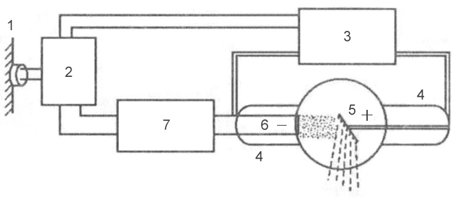 Rice. 3 - block diagram of a typical X-ray machine
 1 - network;
2 - autotransformer;
3 - step-up transformer;
4 - X-ray tube;
5 - anode;
6 - cathode;
7 - step-down transformer.
Mechanism of X-ray generation
X-rays are formed at the moment of collision of a stream of accelerated electrons with the anode substance. When electrons interact with a target, 99% of them kinetic energy turns into thermal energy and only 1% - into x-ray radiation.
An X-ray tube consists of a glass cylinder into which 2 electrodes are soldered: a cathode and an anode. The air has been pumped out of the glass balloon: the movement of electrons from the cathode to the anode is possible only under conditions of relative vacuum (10 -7 –10 -8 mm Hg). The cathode has a filament, which is a tightly twisted tungsten spiral. When submitting electric current Electron emission occurs on the filament, in which electrons are separated from the filament and form an electron cloud near the cathode. This cloud is concentrated at the focusing cup of the cathode, which sets the direction of electron motion. The cup is a small depression in the cathode. The anode, in turn, contains a tungsten metal plate onto which electrons are focused - this is where X-rays are produced.
Rice. 4 - device x-ray tube:
A - cathode; 
B - anode;
B - tungsten filament;
G - focusing cup of the cathode;
D - flow of accelerated electrons;
E - tungsten target;
F - glass flask;
Z - window made of beryllium;
And - formed x-rays;
K - aluminum filter.
There are 2 transformers connected to the electronic tube: a step-down and a step-up. A step-down transformer heats the tungsten coil with low voltage (5-15 volts), resulting in electron emission. A step-up, or high-voltage, transformer fits directly to the cathode and anode, which are supplied with a voltage of 20–140 kilovolts. Both transformers are placed in the high-voltage block of the X-ray machine, which is filled with transformer oil, which ensures cooling of the transformers and their reliable insulation.
After an electron cloud has been formed using a step-down transformer, the step-up transformer is turned on, and a high-voltage voltage is applied to both poles of the electrical circuit: a positive pulse to the anode, and a negative pulse to the cathode. Negatively charged electrons are repelled from the negatively charged cathode and tend to the positively charged anode - due to this potential difference, high speed movement - 100 thousand km/s. At this speed, electrons bombard the tungsten plate of the anode, short-circuiting electrical circuit, resulting in the generation of x-rays and thermal energy.
X-ray radiation is divided into bremsstrahlung and characteristic. Bremsstrahlung occurs due to a sharp slowdown in the speed of electrons emitted by a tungsten helix. Characteristic radiation occurs at the time of perestroika electronic shells atoms. Both of these types are formed in the X-ray tube at the moment of collision of accelerated electrons with atoms of the anode substance. The emission spectrum of an X-ray tube is a superposition of bremsstrahlung and characteristic X-rays.
 Rice. 5 - principle of formation of bremsstrahlung X-ray radiation.
 Rice. 6 - principle of formation of characteristic x-ray radiation.
Basic properties of X-ray radiation
- X-rays are invisible to visual perception.
- X-ray radiation has a high penetrating ability through organs and tissues of a living organism, as well as dense structures inanimate nature, do not transmit visible light rays.
- X-rays cause some to glow chemical compounds, called fluorescence.
- Zinc and cadmium sulfides fluoresce yellow-green,
- Calcium tungstate crystals are violet-blue.
Electromagnetic vibration scale
X-rays have a specific wavelength and vibration frequency. The wavelength (λ) and oscillation frequency (ν) are related by the relation: λ ν = c, where c is the speed of light, rounded to 300,000 km per second. The energy of X-rays is determined by the formula E = h ν, where h is Planck's constant, a universal constant equal to 6.626 10 -34 J⋅s. The wavelength of the rays (λ) is related to their energy (E) by the ratio: λ = 12.4 / E.
X-ray radiation differs from other types of electromagnetic oscillations in wavelength (see table) and quantum energy. The shorter the wavelength, the higher its frequency, energy and penetrating power. The X-ray wavelength is in the range
. By changing the wavelength of X-ray radiation, its penetrating ability can be adjusted. X-rays have a very short wavelength, but a high vibration frequency, and are therefore invisible to the human eye. Due to their enormous energy, quanta have great penetrating power, which is one of the main properties that ensure the use of X-ray radiation in medicine and other sciences.Characteristics of X-ray radiation
Intensity - quantitative characteristic X-ray radiation, which is expressed by the number of rays emitted by the tube per unit time. The intensity of X-ray radiation is measured in milliamps. Comparing it with the intensity of visible light from a conventional incandescent lamp, we can draw an analogy: for example, a 20-watt lamp will shine with one intensity, or strength, and a 200-watt lamp will shine with another, while the quality of the light itself (its spectrum) is the same . The intensity of an X-ray is essentially the amount of it. Each electron creates one or more quanta of radiation at the anode, therefore, the number of X-rays when exposing an object is regulated by changing the number of electrons tending to the anode and the number of interactions of electrons with atoms of the tungsten target, which can be done in two ways:
- By changing the degree of heating of the cathode spiral using a step-down transformer (the number of electrons generated during emission will depend on how hot the tungsten spiral is, and the number of radiation quanta will depend on the number of electrons);
- By changing the magnitude of the high voltage supplied by a step-up transformer to the poles of the tube - the cathode and the anode (the higher the voltage is applied to the poles of the tube, the more kinetic energy the electrons receive, which, due to their energy, can interact with several atoms of the anode substance in turn - see. rice. 5; electrons with low energy will be able to enter into smaller number interactions).
The X-ray intensity (anode current) multiplied by the exposure time (tube operating time) corresponds to the X-ray exposure, which is measured in mAs (milliamperes per second). Exposure is a parameter that, like intensity, characterizes the number of rays emitted by the X-ray tube. The only difference is that the exposure also takes into account the operating time of the tube (for example, if the tube works for 0.01 seconds, then the number of rays will be one, and if 0.02 seconds, then the number of rays will be different - twice more). The radiation exposure is set by the radiologist on the control panel of the X-ray machine, depending on the type of examination, the size of the object being examined and the diagnostic task.
Rigidity - quality characteristic X-ray radiation. It is measured by the magnitude of the high voltage on the tube - in kilovolts. Determines the penetrating power of x-rays. It is regulated by the high voltage supplied to the X-ray tube by a step-up transformer. The higher the potential difference is created at the electrodes of the tube, the more greater strength electrons are repelled from the cathode and rush to the anode, and the stronger their collision with the anode. The stronger their collision, the shorter the wavelength of the resulting X-ray radiation and the higher the penetrating ability of this wave (or the hardness of the radiation, which, like the intensity, is regulated on the control panel by the voltage parameter on the tube - kilovoltage).
λ - wavelength;  Rice. 7 - Dependence of wavelength on wave energy:
E - wave energy  Rice. 8 - The relationship between the voltage on the X-ray tube and the wavelength of the resulting X-ray radiation:
Classification of X-ray tubes
- By purpose
- Diagnostic
- Therapeutic
- For structural analysis
- For translucent
- By design
- By focus
- Single-focus (one spiral on the cathode, and one focal spot on the anode)
- Bifocal (two spirals on the cathode different sizes, and there are two focal spots on the anode)
- By anode type
- Stationary (fixed)
- Rotating
X-rays are used not only for x-ray diagnostic purposes, but also for therapeutic purposes. As noted above, the ability of X-ray radiation to suppress the growth of tumor cells makes it possible to use it in radiation therapy for cancer. In addition to the medical field of application, X-ray radiation has found wide application in engineering, materials science, crystallography, chemistry and biochemistry: for example, it is possible to identify structural defects in various products (rails, welds, etc.) using X-ray radiation. This type of research is called flaw detection. And at airports, train stations and other places mass gathering X-ray television introscopes are actively used for X-ray examination of people hand luggage and luggage for security purposes.
Depending on the type of anode, X-ray tubes vary in design. Due to the fact that 99% of the kinetic energy of electrons is converted into thermal energy, during operation of the tube, significant heating of the anode occurs - the sensitive tungsten target often burns out. The anode is cooled in modern X-ray tubes by rotating it. The rotating anode has the shape of a disk, which distributes heat evenly over its entire surface, preventing local overheating of the tungsten target.
The design of X-ray tubes also differs in terms of focus. The focal spot is the area of the anode where the working X-ray beam is generated. Divided into real focal spot and effective focal spot ( rice. 12). Because the anode is angled, the effective focal spot is smaller than the actual one. Various sizes focal spot are used depending on the size of the image area. The larger the image area, the wider the focal spot must be to cover the entire area of the image. However, a smaller focal spot produces better image clarity. Therefore, when producing small images, a short filament is used and electrons are directed to a small target area of the anode, creating a smaller focal spot.
 Rice. 9 - X-ray tube with a stationary anode.
 Rice. 10 - X-ray tube with a rotating anode.
 Rice. 11 - X-ray tube device with a rotating anode.
 Rice. 12 is a diagram of the formation of a real and effective focal spot.
X-rays are a type of electromagnetic wave, which also includes light rays, radium gamma rays and rays emitted by radio antennas. Electromagnetic waves grouped by their lengths. At the long-wave end of the spectrum, their length ranges from 10 cm to several kilometers. As it decreases, the region of infrared or heat waves begins. The visible light region includes wavelengths (depending on color) from 800 to 400 mm k. The ultraviolet region includes waves from 180 to 10 mm k.
Waves from 15A to 0.03A are characteristic of X-rays. Gamma rays have smaller wavelengths, on the order of 0.001 A radioactive decay. The unit of length angstrom (A) is equal to one hundred millionth of a centimeter.
All these types of radiation differ from one another in the nature of their occurrence and the nature of their interaction with environment. Various properties rays are caused by unequal wavelengths.
Electromagnetic oscillations are also characterized by the amount of quantum energy (a quantum is a separate portion of radiation energy). The shorter the radiation wavelength, the larger value quantum energy.
The laws of propagation of X-rays are similar to the laws of propagation of light. Like light radiation, X-rays, when interacting with the environment, are partially absorbed, partially reflected and scattered. But since the wavelength of X-rays is small and the energy of the quanta is high, they have other properties: 1) penetrate through media various densities- cardboard, wood, animal tissue, etc. The shorter the wavelength and, therefore, the greater the energy of the quanta, the greater the penetrating ability of X-rays. The depth of penetration of X-rays into a particular medium, or the degree of attenuation of the intensity of X-rays when passing through a layer of a particular material, depends not only on the short wavelength or energy of the quanta, but also on the properties of the material: the denser the medium, the more X-rays are absorbed in it rays. For example, a 35 cm thick layer of water attenuates the intensity of the X-ray flux generated at a voltage of 200 kV to the same extent as a 4.75 cm thick layer of iron or 17.23 cm thick concrete;
2) cause glow - luminescence of some chemical compounds. Some substances glow when exposed to X-rays; this glow is called fluorescence. Other substances continue to glow for some time after the X-rays have stopped acting, this glow is called phosphorescence;
3) like visible light, they cause changes in the silver halide compounds that are part of photographic emulsions. In other words, they cause photochemical reactions;
4) cause ionization of neutral atoms and molecules. As a result of ionization, positively and negatively charged particles are formed - ions. The ionized medium becomes a conductor of electric current. This property is used to measure the intensity of rays using a so-called ionization chamber.
At the core biological action X-rays are the phenomenon of ionization.
Rays, which are now called X-rays, were discovered on November 7, 1895 by physicist V. K. Roentgen. The official date of discovery of these rays is December 28, 1895, when Roentgen, after studying the X-rays he discovered, published the first report on their properties.
These X-rays began to be called X-rays on January 23, 1896, when V. K. Roentgen made a public report on X-rays at a meeting of the Physico-Medical Society. At this meeting, it was unanimously decided to call X-rays X-rays.
The nature of X-rays remained little studied for 17 years from the date of their discovery by V.K. Roentgen, although soon after the discovery of these rays the scientist himself and a number of other researchers noted their similarity with visible rays.
The similarity was confirmed by the straightness of propagation and the absence of their deviation in electric and magnetic fields. But, on the other hand, it was not possible to detect either the phenomenon of refraction by a prism, or reflection from mirrors and a number of other properties characteristic of visible light, which has a wave nature.
And only in 1912, first our compatriot, the famous Russian physicist A.I. Lebedev, and then the German physicist Laue, managed to prove that X-rays are of the same nature as rays of visible light, that is, they are electromagnetic waves. Thus, X-rays are the same in nature as radio waves, infrared rays, visible light rays and ultraviolet rays.
The only difference between these rays is that they have different wavelengths of electromagnetic oscillations. Among the above, X-rays have a very short wavelength. Therefore, they required special conditions for the production of experiments to identify refraction or reflection.
The wavelength of X-rays is measured in a very small unit called an angstrom (1 Å = 10–8 cm, that is, one hundred millionth of a centimeter). In practice, diagnostic devices produce rays with a wavelength of 0.1–0.8 Å.
Properties of X-rays
X-rays pass through opaque bodies and objects, such as, for example, paper, matter, wood, human and animal tissue, and even through metals of a certain thickness. Moreover, the shorter the wavelength of radiation, the easier they pass through the listed bodies and objects.
In turn, when these rays pass through bodies and objects with different densities, they are partially absorbed. Dense bodies absorb X-rays more intensely than low-density bodies.
X-rays have the ability to produce visible light in certain chemical substances. For example: barium platinum cyanide crystals, when exposed to X-rays, begin to glow with a bright greenish-yellowish light. The glow continues only at the moment of exposure to x-rays and immediately stops with the cessation of irradiation. Platinum cyanide barium thus fluoresces from the action of X-rays. (This phenomenon was the reason for the discovery of X-rays.)
When illuminated with x-rays, calcium tungstic acid also glows, but with blue light, and the glow of this salt continues for some time even after the irradiation has stopped, i.e. phosphorescent.
The property of causing fluorescence is used to produce transillumination using X-rays. The property of causing phosphorescence in some substances is used to produce x-rays.
X-rays also have the ability to act on the photosensitive layer of photographic plates and films like visible light, causing the decomposition of silver bromide. In other words, these rays have a photo-chemical effect. This circumstance makes it possible to take photographs using X-rays from various parts of the body in humans and animals.
X-rays have a biological effect on the body. Passing through a certain area of the body, they produce corresponding changes in tissues and cells depending on the type of tissue and the amount of rays absorbed by them, i.e. the dose.
This property is used to treat a number of diseases in humans and animals. When exposed to large doses of X-rays, a number of functional and morphological changes occur in the body, and a specific disease arises - radiation sickness .
X-rays, in addition, have the ability to ionize air, that is, split the constituent parts of air into separate, electrically charged particles.
As a result, the air becomes an electrical conductor. This property is used to determine the number of X-rays emitted by an X-ray tube per unit of time using special devices - dosimeters.
Knowing the radiation dose from the X-ray tube is important when X-ray therapy is performed. Without knowing the radiation dose of the tube at the appropriate rigidity, it is impossible to carry out treatment with X-rays, since instead of improving, it is easy to worsen the entire disease process. Improper use of X-rays for treatment can destroy healthy tissue and even cause serious damage throughout the body.